
Morphological Changes in LIM1863 CRC Cells Induced by TGF-β: EMT Assay Analysis
LIM1863 human colorectal cancer (CRC) cells cultured without (A) and with (B) 2 ng/ml TGF-β exhibit significant morphological alterations. In the absence of TGF-β, cells form floating, disorganized multicellular colonies. Conversely, TGF-β exposure leads to adherence to plastic surfaces and a transformation into a monolayer with distinct morphological features indicative of a ‘mesenchymal’ phenotype, including a ‘fibroblastoid’ appearance and a loss of E-cadherin localization. An EMT assay was performed to quantify the area of adherent colonies.

Morphological Changes in LIM1863 CRC Cells Induced by TGF-β: EMT Assay Analysis
E N D
Presentation Transcript
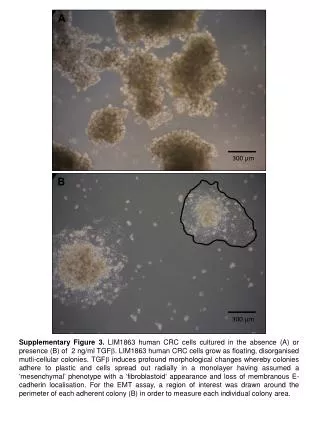
A 300 µm B 300 µm Supplementary Figure 3. LIM1863 human CRC cells cultured in the absence (A) or presence (B) of 2 ng/ml TGFb. LIM1863 human CRC cells grow as floating, disorganised mutli-cellular colonies. TGFbinduces profound morphological changes whereby colonies adhere to plastic and cells spread out radially in a monolayer having assumed a ‘mesenchymal’ phenotype with a ‘fibroblastoid‘ appearance and loss of membranous E-cadherin localisation. For the EMT assay, a region of interest was drawn around the perimeter of each adherent colony (B) in order to measure each individual colony area.