Download
1 / 44
440 likes | 704 Views
HEMOGLOBIN. DR AMINA TARIQ BIOCHEMISTRY. HEME PROTEINS. These are a group of specialized proteins that contain heme and globin . Heme is the prosthetic part and globin is the protein part. 97% is the globin part and the rest 3% is the heme part. GLOBULAR HEME PROTEINS.

E N D
HEMOGLOBIN DR AMINA TARIQ BIOCHEMISTRY
HEME PROTEINS • These are a group of specialized proteins that contain heme and globin. • Heme is the prosthetic part and globin is the protein part. • 97% is the globin part and the rest 3% is the heme part.
GLOBULAR HEME PROTEINS • Role of heme group is dictated by the environment. • Examples: a. Cytochromes b. Catalase c. Hemoglobin d. Myoglobin
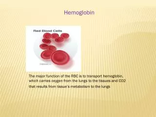
HEMOGLOBIN • Hemoglobin is the protein that carries oxygen from the lungs to the tissues and carries carbon dioxide from the tissues back to the lungs..
The oxygen-carrying protein hemoglobin was discovered in 1840.
Hemoglobin's reversible oxygenation was described a few years later. • In 1959 Max Perutz determined the molecular structure of hemoglobin by X-ray crystallography. This work resulted in his sharing with John Kendrew the 1962 Nobel Prize in Chemistry.
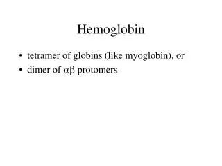
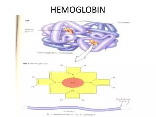
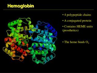
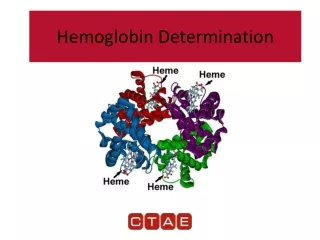
STRUCTURE OF HEMOGLOBIN • Hemoglobin molecule consists of four polypeptide chains: • Two alpha chains, each with 141 amino acids and • Two beta chains, each with 146 amino acids.
Both the α and βglobin chains contain primarily α helix secondary structure with no β sheets.
Each α or βglobin chain folds into 8 α - helical segments (A-H) which, in turn, fold to form globular tertiary structures. • The folded helices form a pocket that holds the working part of each chain, the heme.
The alpha and beta subunits of the globin chains exist in two dimers which are bonded together strongly. • Thus Hb is a tetramer composed of two identical dimers (αβ)1 (αβ)2 .
Quaternary structure of Hb • Polypeptidesof α and β chain are held together by hydrophobic interactions. • Polypeptides between αβ dimers are held by ionic and hydrogen bonds.
PORPHYRIN METABOLISM • Porphyrins are cyclic compounds. • They bind metal ions, mostly Fe2+ or Fe 3+ • The most prevalent metalloporphyrin in humans is Heme. • Heme is the prosthetic group for myoglobin, hemoglobin , cytochromes, catalase and tryptophan pyrrolase.
Heme consists of one ferrous ion in the center of a tetrapyrrole ring of protoporphyrin IX.
Structure of Porphyrins • These are cyclic molecules. • Formed by the linkage of four tetrapyrrole rings, through methenyl bridges. • Structural Features: 1. Side chains- All the porphyrins vary in the nature of their side chains that are attached to their pyrrolerings.e.g.
Uroporphyrin- acetate and propionate • Coproporphyrin- methyl and propionate • Protoporphyrin IX- vinyl, methyl and propionate
2. The side chains can be ordered in four different ways, designated as I- IV. Only Type III porphyrins are physiologically important. They have an asymmetric distribution. e.g. AP, AP,AP, AP- Type I AP, AP, PA, AP- Type III
3. Porphyrinogens : These are the precursors of porphyrins. They are colorless.
STEPS OF SYNTHESIS OF HEME • Major Sites: • Liver (heme proteins- cytochromes)(fluctuating) • Bone marrow (RBC)(constant). • Initial and the last three steps occur in the mitochondria • Intermediate steps in the cytosol. • RBC’s have no mitochondria, unable to synthesize heme.
Glycine + succinylCoA δ-aminolevulinic acid(ALA) Enzyme: Mitochondrial enzyme δ-aminolevulinatesynthase −Hemin, Heme
Reaction requires pyridoxal phosphate as a co- enzyme. • It is the rate limiting step • Inhibited by end product hemin(heme). • Drugs such as phenobarbitol, griseofulvin or hydantoin- increase the activity of ALA synthase.
δ-aminolevulinic acid(ALA) (2 mol condense) Porphobilinogen Enzyme: δ-aminolevulinic acid dehydratase − Lead
Porphobilinogen( 4 molecules condense) Hydroxymethylbilane Enzyme: Hydroxymethylbilanesynthase
Hydroxymethylbilane (ring closure and isomerization) Uroporphyrinogen III Enzyme- Uroporphyrinogen III synthase
Uroporphyrinogen III Coporphyrinogens III Enzyme:Uroporphyrinogendecarboxylase
Coporphyrinogens III Protoporphyrinogen IX Enzyme:CoporphyrinogensOxidase
Protoporphyrinogen IX Protoporphyrin IX Enzyme:Protoporphyrinogenoxidase
Protoporphyrin IX Heme Enzyme:Ferrochelatase
Types in humans In the embryo: • Gower 1 (ζ2ε2) • Gower 2 (α2ε2) • Hemoglobin Portland (ζ2γ2)
In the fetus: • Hemoglobin F (α2γ2)
In adults: • Hemoglobin A (α2β2) - The most common with a normal amount over 95% • Hemoglobin A2 (α2δ2) - δ chain synthesis begins late in the third trimester and in adults, it has a normal range of 1.5-3.5% • Hemoglobin F (α2γ2) - In adults Hemoglobin F is restricted to a limited population of red cells called F-cells. However, the level of Hb F can be elevated in persons with sickle-cell disease.
Hemoglobin C (α2βC2) - Another variant due to a variation in the β-chain gene. This variant causes a mild chronic hemolytic anemia. • Hemoglobin E (α2βE2) - Another variant due to a variation in the β-chain gene. This variant causes a mild chronic hemolytic anemia.
Variant forms which cause disease: • Hemoglobin H (β4) - A variant form of hemoglobin, formed by a tetramer of β chains, which may be present in variants of α thalassemia. • Hemoglobin Barts (γ4) - A variant form of hemoglobin, formed by a tetramer of γ chains, which may be present in variants of α thalassemia.
Hemoglobin S (α2βS2) - A variant form of hemoglobin found in people with sickle cell disease. There is a variation in the β-chain gene, causing a change in the properties of hemoglobin which results in sickling of red blood cells.
Hemoglobin AS - A heterozygous form causing Sickle cell trait with one adult gene and one sickle cell disease gene • Hemoglobin SC disease - Another heterozygous form with one sickle gene and another encoding Hemoglobin C.
In diabetics whose glucose usually runs high, the percent Hb A1c also runs high. Because of the slow rate of Hb A combination with glucose, the Hb A1c percentage is representative of glucose level in the blood averaged over a longer time (the half-life of red blood cells, which is typically 50–55 days).
The levels of glycosylated hemoglobin are tested to monitor the long-term control of the chronic disease of type 2 diabetes mellitus (Type2 DM). Poor control of Type2 DM results in high levels of glycosylated hemoglobin in the red blood cells.
The normal reference range is approximately 4 %–5.9 %. Though difficult to obtain, values less than 7 % are recommended for people with Type 2 DM. Levels greater than 9 % are associated with poor control of diabetes and levels greater than 12 % are associated with very poor control.
Diabetics who keep their glycosylated hemoglobin levels close to 7 % have a much better chance of avoiding the complications that can sometimes accompany diabetes (than those whose levels are 8 % or higher).
Diagnostic uses • Hemoglobin concentration measurement is among the most commonly performed blood tests, usually as part of a complete blood count.
Learning Resources • Lippincott's Biochemistry • Lecture notes