Download
1 / 29
340 likes | 637 Views
IMMUNOGLOBULINS S TRUCTURE AND F UNCTION. BSc Public Health 5th week , 2014. IMMUNOGLOBULINS. Definition

E N D
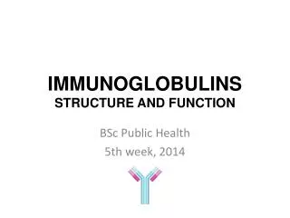
IMMUNOGLOBULINSSTRUCTURE AND FUNCTION BSc Public Health 5th week, 2014
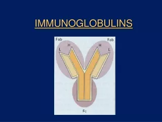
IMMUNOGLOBULINS Definition Glycoprotein molecules that are present on B cells (BCR) or produced by plasma cells (usually referred to as antibodies) in response to an immunogen (antigenthatprovokesimmuneresponse)
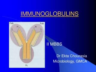
Immunoglobulin STRUCTURE • 2x Heavy chain (light blue) • 2x light chain (dark blue) • Variable regions antigen binding • Constant regions disulfide bond carbohydrate CL VL CH3 CH2 CH1 hinge region VH
Antibody BCR (B cell receptor) mIg sIg Transmembrane domain Associated chains for signaling Cytoplasmic domain SOLUBLE (freely circulating) MEMBRANE BOUND! Antigen bindingeffector functions Produced by plasma cells Antigen recognition B cell activation
ANTIBODY DOMAINS AND THEIR FUNCTIONS Antigen recognition Variable domain Ag Ag Constant domains Effector functions
B CELL ACTIVATION B cell BCR oligomerization results in B cell activation, proliferation and differentiation
Antigen Binding Fragment (Fab) Complement bindingsite Constant fragment (Fc) Binding to Fc receptors on phagocytic cells Placental transfer ANTIGEN BINDING
HYPERVARIABLE REGIONS B celldevelopmentintheredbonemarrowDNA recombination (somaticgenerearrangement) of gene segments encoding variable domains of heavy and light polypeptide chains is responsibleforgeneration of B cellswithhighlyvariablespecificity Light chain CDR2 CDR3 CDR1 Epitope CDR3 CDR1 CDR2 Heavy chain CDR = complementaritydeterminingregion = hypervariableregion
DIFFERENT VARIABLE REGIONS DIFFERENT ANTIGEN-BINDING SITES DIFFERENT SPECIFICITIES
ISOTYPE (CLASS) Sequencevariability of H/L-chainconstantregions • IgG - gamma (γ) heavy chains • IgM - mu (μ) heavy chains • IgA - alpha (α) heavy chains • IgD - delta (δ) heavy chains • IgE - epsilon (ε) heavy chains
MAIN CHARACTERISTICS OF ANTIBODY ISOTYPES IgG1-IgG4 IgA1-IgA2
ANTIBODY PRODUCTION DURING THE PRIMARY AND THE SECONDARY IMMUNE RESPONSES Level of antibodies secondary response against antigen A Primary response against antigen A primary response against antigen B Days napok Antigen A Antigen A and B
EFFECTOR FUNCTIONS OF ANTIBODIES Antibody-mediated immune responses • Fab part: NEUTRALIZATION • Fc part: • OPSONIZATIONfollowedby • opsonizedphagocytosis (macrophage; IgG) • ADCC (NK cell; IgG) • mast cell degranulation (parasite, allergy; IgE) • COMPLEMENT ACTIVATION
Antigenbinding Complement binding site Binding to Fc receptors Placental transfer
OPSONIZED PHAGOCYTOSIS Flagging a pathogen Antigen binding fragment(Fab) binds the pathogenthe Fc region is accessiblefor Fc-receptors of phagocytic cells, facilitating (speeding up)the process of phagocytosis
Opsonization facilitate and acceleratetherecognition of thepathogensbyphagocytes Phagocytes must expressreceptorsfortheopsonins: IgG FcγRI C3b CR1 • Main opsonins: • antibodies • Complementmolecules • Acute-phaseproteins (CRP, SAP)
MAST CELL DEGRANULATION FcεRI + IgE (A) High-affinity FcRs on the surface of the cell bind antibodiesbefore it binds to antigen. (mast cell) (B) Low-affinity FcRs bind multiple Igs that have already bound to a multivalent antigen. (macrophage, NK cell)
Antigenbinding Complement bindingsite Binding to Fc receptors Placental transfer
Antigenbinding Complement binding site Binding to Fc receptors Placental transfer FcRn on the placenta facilitate the transfer of maternal IgG to the body of thefetus
PRODUCTION OF IMMUNOGLOBULINS • IgG transport is so efficient that at birth babies have as high a level of IgG in their plasma as their mothers • These transfers are a form of passive immunization. The babies protection by IgG and IgA is against those pathogen that the mother has mounted • The childrenare most vulnerableduringthefirst year of life (esp.3-12m) whenmaternal IgGshavedisappearedbutthe de novosynthesis is atlowlevel
Pathological consequences of placental transport of IgG (hemolytic disease of the newborn) anti-Rh IgM Passive anti-D IgG
S S S S S S S S S S S S S S S S S S S S S S S S S S S S s s s s s s s s s s s s s s C C C C C C C J J J J J J J C C C C C C C S S S S S S S S S S S S S S C C C C C C C C C C C C C C pIgR and IgA are internalised S S S S S S S S S S S S S S Polymeric Ig receptors are expressed on the basolateral surface of epithelial cells to capture IgA produced in the mucosa C C C C C C C C C C C C C C SECRETORY IgA AND TRANSCYTOSIS ‘Stalk’ of the pIgR is degraded to release IgA containing part of the pIgR (the secretory component) MUC US IgA and pIgR are transported to the apical surface in vesicles Epithelial cell plasmacells located in the submucosa produce dimeric IgA