Download
1 / 20
250 likes | 519 Views
Microscope. The invention of the microscope made it possible for people to discover and learn about cells. A microscope is an instrument that makes small objects look larger. Magnification . The ability to make an object look larger than it is through the bending of light that passes

E N D
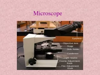
Microscope • The invention of the microscope made it possible for people to discover and learn about cells. • A microscope is an instrument that makes small objects look larger.
Magnification The ability to make an object look larger than it is through the bending of light that passes through lenses.
Resolution • Resolution is the ability to clearly distinguish the individual parts of an object.
ModernMicoscopes • The light microscope. The common light microscope used in the laboratory is called a compound microscope because it contains two types of lenses that function to magnify an object. • The more traditional form of electron microscope is the transmission electron microscope (TEM). To use this instrument, one places ultrathin slices of microorganisms or viruses on a wire grid and then stains them with gold or palladium before viewing. The densely coated parts of the specimen deflect the electron beam, and both dark and light areas show up on the image. • The scanning electron microscope (SEM) is the more contemporary form electron microscope. Although this microscope gives lower magnifications than the TEM, the SEM permits three-dimensional views of microorganisms and other objects. Whole objects are used, and gold or palladium staining is employed.
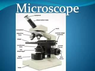
Click on Me Click on Me Click on Me Click on Me Click on Me Click on Me Click on Me Click on Me Click on Me Click on Me Click on Me Click on Me Click on Me Click on Me
#9 Eye Piece/Ocular Lense The part you look at with your eye. Usually 10 X magnification. Click Here to Return to the Main Slide
#10 Arm – Used to safely transport microscope Click Here to Return to the Main Slide
#11 Stage – Slides are placed on this Click Here to Return to the Main Slide
#12 Coarse Adjustment Knob – Used to make large changes in focus. NOTE Never use this when viewing on high power Click Here to Return to the Main Slide
#13 Fine Adjustment Knob Used to small adjustments of focus Click Here to Return to the Main Slide
#14 Base – Used to safely transport the microscope Click Here to Return to the Main Slide
#1 Body Tube – Reflects light up to the viewers eye Click Here to Return to the Main Slide
#2 Nosepiece– Allows for quick change of objectives Click Here to Return to the Main Slide
#3 Low Power Objective – The first lens you use when doing proper microscope work. Usually 4 X Click Here to Return to the Main Slide
#4 Medium Power Objective – The second lens you use when doing proper microscope work. Usually 10 X Click Here to Return to the Main Slide
#5 High Power Objective – The highest magnification used. Usually 43 X. NEVER use the course adjustment when using this lens. Click Here to Return to the Main Slide
#6 Stage Clips – Use to keep the slide in place. Click Here to Return to the Main Slide
#7 Diaphragm – Use to vary the amount of light passing through the slide. Usually it is better if the amount of light is low. Click Here to Return to the Main Slide
#8 Light Source – Sends light up through the diaphragm and through the slide for viewing Click Here to Return to the Main Slide
Rules • Carry MICROSCOPE by arm and base. • Always lower the stage BEFORE you move the objective lenses. • Always have a cover slip over the specimen you are looking at. • If you are looking at cells that are translucent you have to stain them with methylene blue or iodine. • Report any incidents to teacher!