Download
1 / 39
390 likes | 506 Views
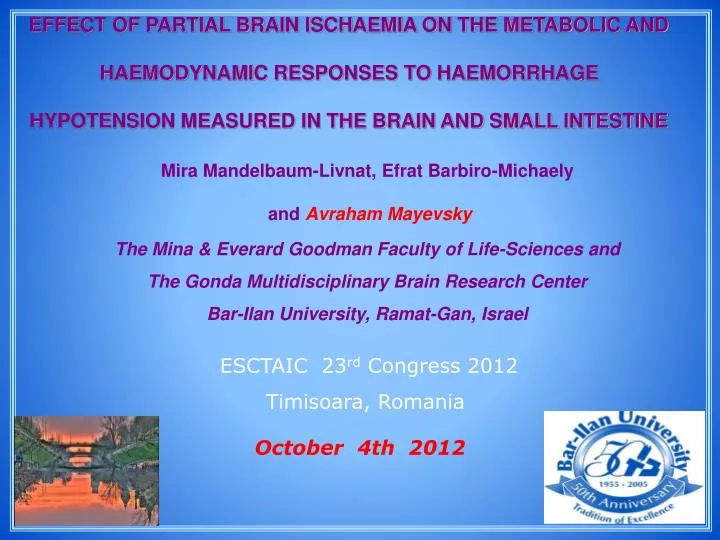
EFFECT OF PARTIAL BRAIN ISCHAEMIA ON THE METABOLIC AND HAEMODYNAMIC RESPONSES TO HAEMORRHAGE HYPOTENSION MEASURED IN THE BRAIN AND SMALL INTESTINE. Mira Mandelbaum-Livnat, Efrat Barbiro-Michaely and Avraham Mayevsky The Mina & Everard Goodman Faculty of Life-Sciences and

E N D
EFFECT OF PARTIAL BRAIN ISCHAEMIA ON THE METABOLIC AND HAEMODYNAMIC RESPONSES TO HAEMORRHAGE HYPOTENSION MEASURED IN THE BRAIN AND SMALL INTESTINE Mira Mandelbaum-Livnat, Efrat Barbiro-Michaely and Avraham Mayevsky The Mina & Everard Goodman Faculty of Life-Sciences and The Gonda Multidisciplinary Brain Research Center Bar-Ilan University, Ramat-Gan, Israel ESCTAIC 23rd Congress 2012 Timisoara, Romania October 4th 2012
Hemorrhage During hemorrhage blood is redistributed in favor of the vital organs and on the expense of the less vital organs. Circulatory blood volume Systemic level Cardiac output Mean Arterial Pressure Blood flow redistribution Sympathetic Nervous System activation O2 delivery less vital organs vital organs Vasoconstriction Autoregulation Tissue level Vessels resistance Vessels resistance Blood flow LDF Blood flow Mitochondrial dysfunction Mitochondrial function preservation NADH Anaerobic metabolism Lactic acidosis ATP cellular level Ionic homeostasis disruption Metabolic acidosis Cell injury Cell death LDF-Laser Doppler Flowmetry
Simultaneous Real Time Monitoring of a Vital Organ - the Brain, and a Less Vital Organ - the Small Intestine, underBody Emergency Metabolic States(BEMS)– a New Approach of Diagnostics Blood Flow Redistribution under BEMS Small Intestine Less Vital Organ Brain Vital Organ
Work hypothesis • During hemorrhage the body suffers from a decrease in oxygen delivery affecting primarily mitochondrial function. • During hemorrhage there is a redistribution of blood from the "less vital organs" (i.e. intestine and skin) to the life preserving circulation of the "vital organs" (i.e. brain and heart). • Monitoring of a “less vital organ” may early detect body emergency metabolic state occurring during hemorrhage.
Importance of research • 50% of the patients with hemorrhagic shock die of substantial blood loss within an hour from the insult. • Another 30% of deaths result from severe internal organ injury during the following 60-120 minutes. • Those who survive their initial injury are at a high risk of developing infection and multi organ failure, which can further lead to death. Early diagnosis of hemodynamic catastrophe and early resuscitation is most important in hemorrhagic shock in order to improve the final outcome.
Comparison of “vital organ” and a “less vital organ” may enable a better understanding of the process occurring on a daily basis in the clinic. • The monitoring of less vital organ during hemorrhage has two important roles: • The early detection of the hemorrhage insult itself. • The early detection of resuscitation end point.
The Multi-Site Multi-Parametric system for monitoring cerebral and intestinal blood flow and mitochondrial NADH
Brain Liver & kidney A1 B1 B2 A2 Testis C C B A D
The development of the monitoring model • In accordance to recent studies reporting about homodynamic differences between two intestinal layers, namely the serosa and mucosa, following hemorrhage we carried out two protocols of short anoxia and epinephrine injection, in order to assure monitoring from both layers and to find out which intestinal location is better for intestinal monitoring.
0 15 45 105 Time (min) 0 15 45 105 Time (min) N2 Epinephrine N2 - death Start N2 N2 N2 - death Start Short anoxia Epinephrine I.V. injection No significant differences between the serosa and mucosa were observed in any of the protocols. Therefore, we have decided to place the intestinal probe on the serosa, since it is less invasive and easier to manipulate.
0 1 2 3 Time (hour) Start control N2 N2 - death Start Normotensive control N=4
Small Intestinal Serosa ) Brain Stop Time (sec) Start Anoxia Intestinal and Brain responses to Anoxia
Small Intestinal Serosa Brain ) ) Start Stop Hypoxia Time (min) Response of Intestine and Brain to Hypoxia
Response of Intestine (gray) and Brain (black) to Hypercapnia
Responses of Intestine (gray) and Brain (black) to Hyperoxia
Small Intestinal Serosa Brain (%) REF (%) NADH (%) TBF ) mmHg ( MAP Intestinal and Brain responses to Epinephrine 10 µg/kg
Small Intestinal Serosa (%) Brain REF (%) NADH (%) TBF ) mmHg ( MAP ** Intestinal and Brain responses to Epinephrine 2-10 µg/kg
(%) Anoxia *** ***
(%) Hypoxia *** ***
Hypercapnia (%) ***
Correlations between NADH & TBF under Epinephrine 2-10mg/kg (I.V)
Hemorrhage Decreased circulatory blood volume Decreased tissue perfusion and O2 delivery Oxygen demand exceeds oxygen supply Hemorrhagic shock
0 1 2 3 4 Time (hour) 15 min Bleeding down to 40 mmHg and maintenance Resuscitation Operation N2 N2 - death Start Sample protocol
0 0 1 1 2 2 3 3 Time (hour) 30 min N2 - death N2 Bleeding Resuscitation Start Averaged amount of shed blood was calculated to be 12±3% of rat’s total blood volume. Uncontrolled hypotension N=9
Controlled hypotension for 15 min Averaged amount of shed blood was calculated to be 31±2% of rat’s total blood volume. N=9
0 0 1 1 2 2 3 3 Time (hour) 30 min N2 - death N2 Bleedingand maintenance Resuscitation Start Averaged amount of shed blood was calculated to be 40±1.5% of rat’s total blood volume. Controlled hypotension for 30 min
Comparison between various models of hypotension Controlled hypotension underNormal cerebral perfusionversusPartial cerebral ischemia
0 24 25 26 27 28 Time (hour) Startcontrol N2 N2 - death Start Bilateral carotid occlusion Operation Partial cerebral ischemia - control group (no hemorrhage) Bilateral carotid occlusion (BCO) is an animal model of arteriosclerosis, which is considered to be the leading cause of mortality in industrialized countries.
0 24 25 26 27 28 Time (hour) 15 min N2 N2 - death Start Resuscitation Bilateral carotid occlusion Operation Bleeding and maintenance Controlled hypotension for 15 min under partial cerebral ischemia
Comparison between the two organs in both experimental groups Brain Intestine The differences between the two models are manifested mainly by the cerebral responses.
Under normal conditions The early signs of the hemorrhage insult were detected in the intestine. The initial deterioration of the intestine, following resuscitation, was not accompanied by a deterioration of MAP. In all of the models the intestine suffered from irreversible damage, whereas the brain remained protected. NADH responses to hemorrhage were higher in the intestine compared to the brain. Under partial cerebral ischemia The response of the ischemic brain, to hemorrhage, was very similar to the intestinal response. The cerebral tissue was suffering from extensive reduction of blood flow. The combination of partial cerebral ischemia and hemorrhagic hypotension blurs the differences between the brain and the small intestine. Conclusions
Conclusions • The intestine may serve as a surrogate organ for monitoring under hemorrhagic insults, due to its capability for early detection of whole body deterioration. • The monitoring of mitochondrial NADH redox state can be used as an indicator of different hemorrhage stress severities. The application of the Multi-site Multi-parametric monitoring system is clearly advantageous under hemorrhagic hypotension.