Download
1 / 17
210 likes | 919 Views
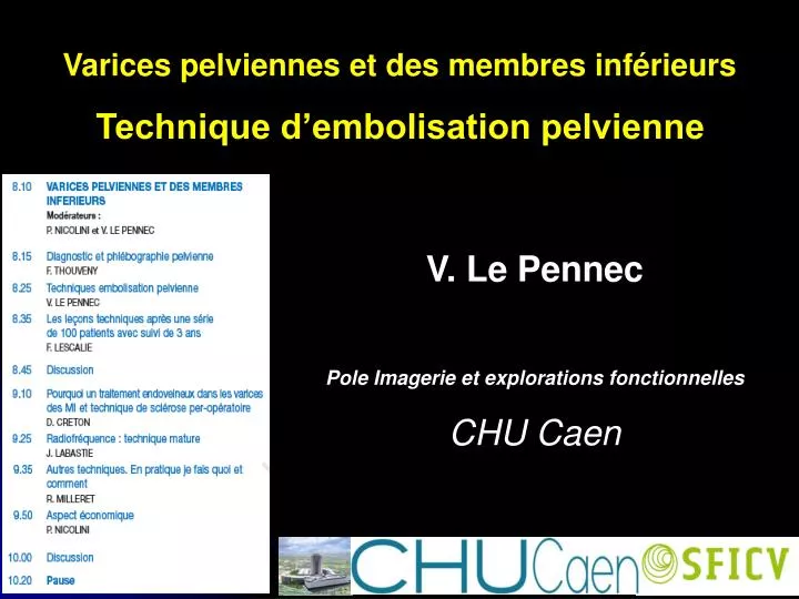
Varices pelviennes et des membres inférieurs Technique d’embolisation pelvienne. V. Le Pennec Pole Imagerie et explorations fonctionnelles CHU Caen. L’ENVIRONNEMENT ENDOVASCULAIRE. Recommandations SFICV – SIR Table télécommandée, soustraction Dosimétrie, radioprotection

E N D
Varices pelviennes et des membres inférieurs Technique d’embolisation pelvienne V. Le Pennec Pole Imagerie et explorations fonctionnelles CHU Caen
L’ENVIRONNEMENT ENDOVASCULAIRE • Recommandations SFICV – SIR • Table télécommandée, soustraction • Dosimétrie, radioprotection • Opérateur expérimenté • Matériel adapté, miniaturisé • Embols connus
LE PREALABLE AU TRAITEMENT • Consultation pré interventionnelle • Phlébographie pelvienne = Gold standard • Cartographies veineuses anatomique et hémodynamique : • Retours veineux rénal et iliocave gauches • Gradient de pression réno-cave • Points de fuite pelviens • Examen indispensable avant tout traitement endovasculaire : • Durée de l’examen trop longue • Remplissage vésical, quantité de PDCI • Corrélation radio-clinique, information éclairée de la patiente
LES PRINCIPES DU TRAITEMENT • Occlusion complète et définitive : • des points de fuite pelviens • de l’alimentation de l’hyperpression veineuse pelvienne • Hospitalisation de jour • Gestion de la douleur post embolisation (AINS) • Agents embolisants
LES COMPLICATIONS • Douleur pelvienne ++++ • Spasme • Extravasation
Récidive (recanalisation de la veine embolisée, afférent de contournement)
LES RESULTATS • Première description d’une embolisation veineuse pelvienne par Edwards en 1993 • 21 publications • 15/21 ont moins de 50 patientes • 131 patientes , Kim JVIR 2006 • Suivi 6 – 44 mois • 9 migrations de matériel, 7 extravasations
Sucès technique > 90% • Succès clinique 70-83% • Dégradation 4% • 5% de récidives Venbrux. Jvir 2002, Kim JVIR 2006, Creton 2007
L’étiologie pelvienne n’est pas évoquée dans la récidive de varices des MI après traitement chirurgical 2B
CONCLUSION • Démarche anatomo-physiologique • Contexte clinique ++++ • Phlébographie pelvienne rétrograde sélective diagnostique • Traitement complet et définitif de l’hyperpression veineuse pelvienne et des points de fuites


![[Radiography] Technique - Exposure Factors](https://cdn0.slideserve.com/546181/radiography-technique-exposure-factors-dt.jpg)