The Forearm 2
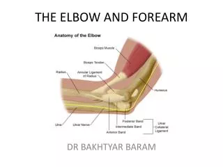
The Forearm 2. Contents of the Lateral Fascial Compartment of the Forearm. The lateral fascial compartment may be regarded as part of the posterior fascial compartment Muscles: Brachioradialis and extensor carpi radialis longus Blood supply: Radial and brachial arteries


The Forearm 2
E N D
Presentation Transcript
Contents of the Lateral Fascial Compartment of the Forearm • The lateral fascial compartment may be regarded as part of the posterior fascial compartment • Muscles: Brachioradialis and extensor carpi radialis longus • Blood supply: Radial and brachial arteries • Nerve supply to the muscles: Radial nerve
Muscles of the Lateral Fascial Compartment of the Forearm • 1. Brachioradialis is ant to elbow joint, acts as an accessory flexor of this joint. • 2. Extensor carpiradialislongus • A:Extends and abducts wrist • Origin • Both of them originates from the Lateral supracondylar ridge of humerus • Insertion • Brachioradialis Base of styloid process of radius • Extensor carpiradialislongus Posterior surface of base of second metacarpal bone • Nerve Supply: • Radial nerve before it divides into superficial and deep branches • The arterial supply is derived from branches of the radial and brachial arteries
Radial Nerve • The radial nerve pierces the lateral intermuscular septum in the lower part of the arm and passes forward into the cubitalfossa • It then passes downward in front of the lateral epicondyle of the humerus, lying between the brachialis on the medial side and the brachioradialis and extensor carpiradialislongus on the lateral side • At the level of the lateral epicondyle, it divides into superficial and deep branches
Branches • Muscular branches to the brachioradialis, to the extensor carpi radialis longus, and a small branch to the lateral part of the brachialis muscle • Articular branches to the elbow joint • Deep branch of the radial nerve. This winds around the neck of the radius, within the supinator muscle and enters the posterior compartment of the forearm • Superficial branch of the radial nerve
Superficial Branch of the Radial Nerve • The superficial branch of the radial nerve is the direct continuation of the nerve after its main stem has given off its deep branch in front of the lateral epicondyle of the humerus • It runs down under cover of the brachioradialis muscle on the lateral side of the radial artery • In the distal part of the forearm, it leaves the artery and passes backward under the tendon of the brachioradialis • It reaches the posterior surface of the wrist, where it divides into terminal branches that supply the skin on the lateral two thirds of the posterior surface of the hand • and the posterior surface over the proximal phalanges of the lateral three and a half fingers. • The area of skin supplied by the nerve on the dorsum of the hand is variable
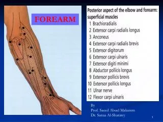
Contents of the Posterior Fascial Compartment of the Forearm • Muscles: • The superficial group includes the • extensor carpiradialisbrevis, • extensor digitorum, • extensor digitiminimi, • extensor carpiulnaris, • and anconeus. • These muscles possess a common tendon of origin, which is attached to the lateral epicondyle of the humerus • The deep group includes • the supinator, • abductor pollicislongus, • extensor pollicisbrevis, • extensor pollicislongus, • and extensor indicis. • Blood supply: Posterior and anterior interosseous arteries • Nerve supply to the muscles: Deep branch of the radial nerve (posterior interosseous nerve).
EXTENSOR CARPI RADIALIS BREVIS • O:Lateral epicondyle of humerus. Radial collaerallig of elbow • I: Dorsal surfaces of bases of 2nd and 3rd metacarpals . • Lies deep to extensor carpiradialislongus. • A:Extends and abducts wrist. • NS: Posterior interosseous nerve.
EXTENSOR DIGITORUM • O: Lateral epicondyle of humerus • Forms four tendons, each passes into a finger • I: Base of dorsal surfaces of middle and distal phalanges. • A:Extends all joints of hand • NS: Posterior interosseous nerve.
EXTENSOR DIGITI MINIMI • An accessory extensor of little finger, medial to extensor digitorum . • O: Lateral epicondyle of humerus • I: Dorsal digital expansion of little finger along with the tendon of extensor digitorum. • A:Extends MCP &IP joints of little finger • NS: Posterior interosseous nerve
EXTENSOR CARPI ULNARIS • Lies medial to the extensor digitiminimi • O: Lateral epicondyle of the humerus Posterior border of ulna by an aponeurosis • I: Medial side of the base of 5th metacarpal . • A: Extends and adducts the wrist • NS: Posterior interosseous nerve
ANCONEUS • Small triangular muscle behind the cubital joint. • O:Post surface of lat epicondyle of humerus. • I: Lat aspect of Olecranon Proximal 1/4th of post surface of ulna. • A: Weak extensor of elbow • NS: Radial Nerve
SUPINATOR • Two heads • O: Superficial head: lat epicondyle of humerus, radial collateral lig of elbow annular lig, • Deep head : supinator crest of ulna,post part of triangular area in front of it • I: Lat surface of prox 1/3 of radius • A: Supinates the forearm and hand • NS: Posterior interroseous nerve.
EXTENSOR CARPI RADIALIS BREVIS • O:Lateral epicondyle of humerus. Radial collaerallig of elbow • I: Dorsal surfaces of bases of 2nd and 3rd metacarpals . • Lies deep to extensor carpiradialislongus. • A:Extends and abducts wrist. • NS: Posterior interosseous nerve.
EXTENSOR DIGITORUM • O: Lateral epicondyle of humerus • Forms four tendons, each passes into a finger • I: Base of dorsal surfaces of middle and distal phalanges. • A:Extends all joints of hand • NS: Posterior interosseous nerve.
EXTENSOR DIGITI MINIMI • An accessory extensor of little finger, medial to extensor digitorum . • O: Lateral epicondyle of humerus • I: Dorsal digital expansion of little finger along with the tendon of extensor digitorum. • A:Extends MCP &IP joints of little finger • NS: Posterior interosseous nerve
EXTENSOR CARPI ULNARIS • Lies medial to the extensor digitiminimi • O: Lateral epicondyle of the humerus Posterior border of ulna by an aponeurosis • I: Medial side of the base of 5th metacarpal . • A: Extends and adducts the wrist • NS: Posterior interosseous nerve
ABDUCTOR POLLICIS LONGUS • O:Prox post surfaces of the radius and ulna, interosseousmemb. • I: Radial side of base of 1st metacarpal . Trapezium • A: Abd and ext of thumb at carpometacarpal joint • NS: Posterior interroseous nerve
EXTENSOR POLLICIS BREVIS • O: Post surface of radius, interosseousmemb. • I:Dorsal surface of base of prox phalanx of thumb. • A:Extends prox phalanx and metacarpal of thumb. • NS: Posterior interroseous nerve.
EXTENSOR POLLICIS LONGUS • O: Posterior surface of ulna interosseousmemb. • I:Dorsal surface of distal phalanx of the thumb • A:Extends all joints of the thumb • NS: Posterior interosseous nerve.
EXTENSOR INDICES • O: Posterior surface of ulna distal to extensor pollicislongus, interosseousmemb . • I:Ulnar side of tendon of extensor digitorum for index finger. • A:Extension of Index finger. • NS: Posterior interosseous nerve
Arteries of the Posterior Fascial Compartment of the Forearm • The anterior and posterior interosseous arteries arise from the common interosseous artery, a branch of the ulnar artery • They pass downward on the anterior and posterior surfaces of the interosseous membrane, respectively, and supply the adjoining muscles and bones. • They end by taking part in the anastomosis around the wrist joint.
Deep Branch of the Radial Nerve • The deep branch arises from the radial nerve in front of the lateral epicondyle of the humerus in the cubitalfossa • pierces the supinator and winds around the lateral aspect of the neck of the radius in the substance of the muscle to reach the posterior compartment of the forearm. • The nerve descends in the interval between the superficial and deep groups of muscles • It eventually reaches the posterior surface of the wrist joint. • Branches : • Muscular branches • Articular branches to the wrist and carpal joints
Extensor Retinaculum • The extensor retinaculum is a thickening of deep fascia that stretches across the back of the wrist and holds the long extensor tendons in position • converts the grooves on the posterior surface of the distal ends of the radius and ulna into six separate tunnels for the passage of the long extensor tendons • Each tunnel is lined with a synovial sheath, which extends above and below the retinaculum on the tendons • The tunnels are separated from one another by fibrous septa that pass from the deep surface of the retinaculum to the bones • The retinaculum is attached medially to the pisiform bone and the hook of the hamate and laterally to the distal end of the radius • The upper and lower borders of the retinaculum are continuous with the deep fascia of the forearm and hand, respectively.
Carpal Tunnel • The bones of the hand and the flexor retinaculum form the carpal tunnel • The median nerve lies in a restricted space between the tendons of the flexor digitorum superficialis and the flexor carpi radialis muscles
Structures on the Anterior Aspect of the Wrist • The following structures pass superficial to the flexor retinaculum from medial to lateral • Flexor carpiulnaris tendon, ending on the pisiform bone • Ulnar nerve lies lateral to the pisiform bone. • Ulnar artery lies lateral to the ulnar nerve. • Palmarcutaneous branch of the ulnar nerve • Palmaris longus tendon (if present), passing to its insertion into the flexor retinaculum and the palmaraponeurosis • Palmarcutaneous branch of the median nerve
The following structures pass beneath the flexor retinaculum from medial to lateral • Flexor digitorumsuperficialis tendons and, posterior to these, the tendons of the flexor digitorumprofundus; both groups of tendons share a common synovial sheath. • Median nerve • Flexor pollicislongus tendon surrounded by a synovial sheath • Flexor carpiradialis tendon going through a split in the flexor retinaculum. The tendon is surrounded by a synovial sheath.
Structures on the Posterior Aspect of the Wrist • The following structures pass superficial to the extensor retinaculum from medial to lateral • Dorsal (posterior) cutaneous branch of the ulnar nerve • Basilic vein • Cephalic vein • Superficial branch of the radial nerve
Beneath the extensor retinaculum, fibrous septa pass to the underlying radius and ulna and form six compartments that contain the tendons of the extensor muscles • Each compartment is provided with a synovial sheath, which extends above and below the retinaculum • The following structures pass beneath the extensor retinaculum from medial to lateral • 1. Extensor carpiulnaris tendon which grooves the posterior aspect of the head of the ulna (num 6 on the figure) • 2. Extensor digitiminimi tendon situated posterior to the distal radioulnar joint. • 3. Extensor digitorum and extensor indicis tendons share a common synovial sheath and are situated on the lateral part of the posterior surface of the radius. • 4. Extensor pollicislongus tendon winds around the medial side of the dorsal tubercle of the radius.
5. Extensor carpiradialislongus and brevis tendons share a common synovial sheath and are situated on the lateral part of the posterior surface of the radius. • 6. Abductor pollicislongus and the extensor pollicisbrevis tendons have separate synovial sheaths but share a common compartment. • The radial artery reaches the back of the hand by passing between the lateral collateral ligament of the wrist joint and the tendons of the abductor pollicislongus and extensor pollicisbrevis
EXTENSOR RETINACULUM • Starting from lateral side. COMPARTMENT STRUCTURES I Abductor pollicislongus Extensor pollicisbrevis II Extensor carpiradialislongus Extensor carpiradialisbrevis III Extensor pollicislongus. IV Extensor digitorium Extensor indicis Post interrosseous nerve Ant interrosseous artery V Extensor digitiminimi. VI Extensor carpiulnaris