Download
1 / 26
270 likes | 381 Views
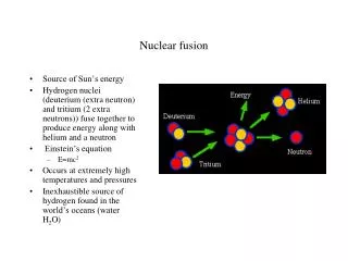
FUSION OF BRUSHLET AND WAVELET DENOISING METHODS FOR NUCLEAR IMAGES. Elsa Angelini 1 , Yinpeng Jin 1 , Peter Esser 2 , R. Van Heertum 2 , Andrew Laine 1 1 Department of Biomedical Engineering 2 Department of Radiology Columbia University, New York, NY, USA. ISBI 2004 Washington, DC

E N D
FUSION OF BRUSHLET AND WAVELET DENOISING METHODS FOR NUCLEAR IMAGES Elsa Angelini1, Yinpeng Jin1, Peter Esser2, R. Van Heertum2, Andrew Laine1 1 Department of Biomedical Engineering 2 Department of Radiology Columbia University, New York, NY, USA ISBI 2004 Washington, DC April 17, 2004
Sample PET and SPECT Images SPECT Liver PET Brain
Previous Work on Multi-Scale Processing of PET and SPECT • Local reconstruction to improve spatial resolution within a region of interest • T. Olson and J. De Stefano, "Wavelet Localization of the Radon Transform." IEEE Trans. Image Processing, vol. 42, pp. 2055-2067, 1994. • F. Rashid-Farrokhi, K. Liu, C. Berenstein, and D. Walnut, "Wavelet-based Multiresolution Local Tomography." IEEE Trans. Image Processing, vol. 22, pp. 1412-1430, 1997. • S. Zhao, G. Wang, and J. Hsieh, "Wavelet Sampling and Localization Schemes for the Radon Transform in Two Dimensions." SIAM Journal on Applied Mathematics, vol. 57, pp. 1749-1762, 1997. • M. Bottema, B. Morean, and S. Suorova, "An Application of Wavelets in Tomography." Digital Signal Processing, vol. 8, pp. 244-254, 1998. • W. Maldych, "Tomography, Approximate Reconstructions, and Continuous Wavelet Transforms." Journal of Applied Computation and Harmonic Analysis, vol. 7, pp. 54-100, 1999. • Accelerating implementation of the traditional FBP algorithm • A. Delaney and Y. Bresler, "Multi-resolution Tomographic Reconstruction Using Wavelets." IEEE Trans. Image Processing, vol. 4, pp. 799-813, 1995. • L. Blanc-Feraud, P. Charbonnier, P. Lobel, and M. Barlaud, "A Fast Tomographic Reconstruction Algorithm in the 2-D Wavelet Transform Domain." IEEE International Conference on Acoustics, Speech and Signal Processing, pp. 305-308, 1994. • Post-filtering or regularization/constraints in tomographic reconstruction • E. Kolaczyk, "A Wavelet Shrinkage Approach to Tomographic Image Reconstruction." Journal of American Statistics Association, vol. 91, pp. 1079-1090, 1996. • N. Lee and B. Lucier, "Wavelet Methods for Inverting the Radon Transform with Noisy Data." IEEE Trans. Image Processing, vol. 10 (1), pp. 79-94, 2001. • J. Lin, A. Laine, and S. Bergmann, "Improving PET-based Methods Using the Wavelet Transform for Positron Emission Tomography." IEEE Trans. Biomedical Engineering, vol. 48, pp. 202-212, 2001. • J. Kalifa, A. Laine, and P. Esser, "Regularization in Tomographic Reconstruction Using Thresholding Estimators." IEEE Trans. Medical Imaging, vol. 22 (3), pp. 351-359, 2003.
Image De-Noising in PET and SPECT Texture-Based De-noising (Brushlet Analysis) Image Fusion Edge-Based De-noising (3D Wavelet Modulus Analysis) • Directional and Textural Noise SPECT PET • Over-Smooth • Structure Edges
Image De-Noising in PET and SPECT Texture-Based De-noising (Brushlet Analysis) Image Fusion Edge-Based De-noising (3D Wavelet Modulus Analysis) • Directional and Textural Noise SPECT PET • Over-Smoothed • Structure Edges
2D Analysis Function Fourier Tiling to construct expansion basis Brushlet and Textural Analysis Brushlets Basis Functions Expansion Reconstruction
Brushlet and Textural Analysis Advantages of Brushlets Analysis • Compact representation of textured signals. • Adaptive tiling of frequency plane. • Adaptive directional selectivity. • Fast implementation with folding operators and FFT. • Orthogonal basis.
De-noising with Brushlet Basis Functions(ISBI 02) Data Mean Regions Variance Map frequency Noise s Brushlet and Textural Analysis Isolate oriented textures via thresholding • Minimax threshold level based on noise variance, estimated in the background. • Spatial adaptivity of thresholding for 3 types of regions: texture, smooth, edges [Vetterli]. • De-noising via hard thresholding of low frequency coefficients.
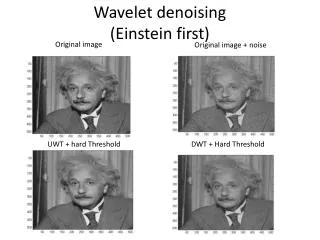
Examples of Brushlet De-Noising: SPECT Brain Data Brushlet and Textural Analysis Original Denoised
Image De-Noising in PET and SPECT Texture-Based De-noising (Brushlet Analysis) Image Fusion Edge-Based De-noising. (3D Wavelet Modulus Analysis) • Directional and Textural Noise SPECT PET • Over-Smoothed • Structure Edges
Edge-Based De-noising Wavelet and Edge De-noising • 3D Dyadic Wavelet Thresholding. • Feature selection based on spatial orientation of contours in three dimensions. • Cross-Scale Regularization(MICCAI’ 03) • Explore correlations of signal features across spatial-frequency scales. • Effective signal recovering from noise-dominated multi-scale expansions.
3D Dyadic Wavelets and Wavelet Modulus DC m,1 m,2 m,3 Input Data Wavelet Coefficients 3D: N,1 N,2 N,3 Wavelet Modulus in 3D: Wavelet Edge De-noising
Traditional* Dyadic Wavelet Thresholding (3D) Wavelet Decomposition Wavelet Reconstruction Threshold Threshold Threshold Input Image Enhanced (Denoised) Image Threshold Threshold Threshold DC Wavelet Edge De-noising * [Mallat 92]
Dyadic Wavelet Modulus Thresholding (3D) Wavelet Decomposition Wavelet Reconstruction Modulus Thresholding Enhanced (Denoised) Image Modulus Thresholding DC Wavelet Voxel De-noising Input Image
Cross-scale Regularization (CSR) Wavelet Edge De-noising Wavelet Modulus at Expansion Levels 1 and 2 - “Pre-processing” of higher level sub-bands: • De-correlation of noise in spatial-frequency expansion Level 1 Level 2 Input Image + x - “Windowed” Normalization - Avoid attenuation of weak edges - 50% Max rule (brain, liver data) - 70% Max rule (bone data) N “Regularization Map”
Comparison: CSR vs. Soft Threshold Wavelet Edge De-noising Input Data CSR De-noising Soft Thresholding
Image De-Noising in PET and SPECT Texture-Based De-noising (Brushlet Analysis) Edge-Based De-noising. (3D Wavelet Modulus Analysis) • Directional and Textural Noise. SPECT PET Image Fusion • Over-Smoothed Structure Edges.
Multi-Scale Image Fusion • Fusion:To combine different or incomplete representations into a unified form with integrated information. • Motivation of fusion in the context of denoising: • Brushlet analysis provides better enhancement of “harmonic textures”, representing physiological activities inside target organs. • Wavelet modulus thresholding provides better enhancement of “anatomicaledges”, or delineation of anatomical structures of clinical interest. • Both types of information are important for accurate diagnostic decisions and image interpretation.
Multi-Scale Image Fusion Fusion Process A B Wavelet Expansion Wavelet Expansion A1 A2 A3 B1 B2 B3 Fusion Rule: Fi(x,y,z) = Max(Ai(x,y,z), Bi(x,y,z)) F1 F2 F3 Wavelet Reconstruction F De-noised Data Sets Both [A] and [B] expanded and reconstructed with 3D Dyadic Transform
Multi-Scale Image Fusion Example: Fusion of coefficient features at the most detailed expansion level. Wavelet Modulus De-Noising Brushlet De-Noising Fused Image Features
Multi-Scale Image Fusion Brushlet De-Noising Input Data Image Fusion Result Wavelet Modulus De-Noising Example Cases: Fusion of denoised images
Multi-Scale Image Fusion Brushlet De-Noising For comparison: Reconstructed Using OSEM Wavelet Modulus De-Noising Example: Fusion of denoised images Input Data: Clinical PET Brain Reconstructed Using FBP Image Fusion Result
Multi-Scale Image Fusion Preliminary Clinical Evaluation • Brushlet de-noising: • Beneficial for enhancing “harmonic activity”, e.g. anatomical or physiological variations within the target organs. • Wavelet modulus analysis with cross-scale regularization: • Beneficial for enhancing “anatomical edges”, with a better definition and delineation of the organ contours. • Fused images: • Effectively combined important features from both processed images, without introducing artifacts. • When compared to OSEM reconstructions, provided significantly improved image quality in terms of both lower noise level and improved contrast for key anatomical and physiological features.
Conclusion • Multi-scale fusion of two expansions • Selected predominant wavelet coefficient modulus from distinct de-noising expansions. • Effective integration of de-noising methods for enhancement of anatomical and physiological features. • Potential improvements of the method • Preservation of the linearity of the nuclear measures. • Refinement of fusion rule. • Further evaluation studies • Clinical phantom data. • Clinical data with pathological ground truth.
References • Y. Jin, E. Angelini, P. Esser, and A. Laine, "De-noising SPECT/PET images using cross-scale regularization," MICCAI, pp. 32-40, Montreal, Canada, 2003. • F. Meyer and R. R. Coifman, "Brushlets: A tool for directional image analysis and image compression," Applied and Computational Harmonic Analysis, vol. 4, No. 1, pp. 147-187, 1997. • E. D. Angelini, J. Kalifa, and A. F. Laine, "Harmonic multiresolution estimators for denoising and regularization of SPECT-PET data," International Symposium on Biomedical Imaging, pp. 697-700, Washington, D.C., USA, 2002. • S. Mallat and S. Zhong, "Signal characterization from multiscale edges," 10th International Conference on Pattern Recognition, pp. 891-896, Atlantic City, NJ, USA, 1990. • E. Angelini, A. Laine, S. Takuma, J. Holmes, and S. Homma, "LV volume quantification via spatio-temporal analysis of real-time 3D echocardiography," IEEE Transactions on Medical Imaging, vol. 20, No. 6, pp. 457-469, 2001. • S. G. Chang, B. Yu, and M. Vetterli, "Spatially adaptive wavelet thresholding with context modeling for image denoising," IEEE International Conference on Image Processing, pp. 535 -539, Chicago, IL, USA, 1998. • S. G. Nikolov, D. R. Bull, C. N. Canagarajah, M. Halliwell, and P. N. T. Wells, "Fusion of 2-D images using their multiscale edges," IEEE International Conference on Pattern Recognition, pp. 41-44, Barcelona, Spain, 2000. • I. Koren, A. Laine, and F. Taylor, "Image fusion using steerable dyadic wavelet transform," IEEE International Conference on Image Processing, pp. 232-235, Washington, D.C., USA, 1995.
Acknowledgements This study was supported in part by Siemens Medical Solutions, Inc.