Download
1 / 78
820 likes | 1.1k Views
Common Problems of the Aging Athlete. Brian L. Badman M.D. Orthopedics and Sports Medicine of Indiana Hendricks Regional Health. Rotator Cuff Injuries. Rotator Cuff Anatomy. 4 muscles/tendons Supraspinatus Infraspinatus Teres Minor Subscapularis. Rotator Cuff Function.

E N D
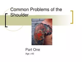
Common Problems of the Aging Athlete Brian L. Badman M.D. Orthopedics and Sports Medicine of Indiana Hendricks Regional Health
Rotator Cuff Anatomy • 4 muscles/tendons • Supraspinatus • Infraspinatus • Teres Minor • Subscapularis
Rotator Cuff Function • Shoulder rotation • Arm elevation • Helps keep humeral head within shoulder socket
Rotator Cuff Injuries • The position of injury for the rotator cuff is overhead WHY?
Rotator Cuff Injuries • Overhead Position • Impingement between humerus and acromion • Least efficient/weakest position for cuff • Poor blood supply to tendons
Rotator Cuff Injuries • What causes the injury? • Trauma • Fall on an outstretched hand • Arm forcefully pulled to side or downward • Overuse • Repetitive lifting, loading
Traumatic Causes • Tear of Tendons • Partial • Complete
Overuse Causes Chronic Inflammation Bursitis Fraying Gradual progression of tear ONCE TEARING STARTS IT IS EASIER TO PROGRESS
Why Does it Worsen? Weakened cuff cannot protect itself Space between acromion and humerus narrows Impingement worsens Tear progresses
Symptoms • Pain in deltoid region • Upper outer arm • Pain worse with overhead activities • Night pain • Pain with exertion • Lifting
Symptoms • MUST ALWAYS EVALUATE NECK AS SYMPTOMS FROM A HERNIATED DISK CAUSING NERVE IMPINGEMENT ARE SIMILIAR
Evaluation • History • Physical Examination • MRI
Evaluation • History • Trauma, location, provocation of symptoms • Exam • Pain with isolated cuff testing • Impingement signs • Weakness
Evaluation • MRI • NEED HIGH QUALITY MAGNET! • Insurance will pay the same so might as well insist on best quality study • Your doctor may not be aware that this is important so stress the need for a CLOSED MRIunless you are claustrophobic • In-office • Open MRI POOR QUALITY STUDIES
Treatment • If have a partial tear or cuff just inflamed (bursitis, tendonitis) • Physical Therapy • NSAIDS (Motrin, alleve, advil, celebrex) • +/- Steroid Injection • Rest • Avoid overhead activities and lifting It may take several months for pain and inflammation to resolve
Treatment • Complete Tear • In active individuals surgery is generally indicated • Rotator cuff will not heal itself • Results are better if a traumatic tear is fixed acutely (2-3 weeks) • Takes 12 weeks for repair to heal to bone • Anticipate a year to recover • 3 monthsbegin overhead activities • 6 monthsMay resume most activities as long as motion is good
Postop Rehab • Because the cuff heals slowly (12 weeks minimum) the repair is easily damaged if stressed too soon after surgery • Slow progression with motion and strengthening is necessary • May result in stiffness • Usually resolves but may take entire year to return
Goals of Surgery • Repair tear • Alleviate pain • Maintain full ROM • Maintain strength
Surgery • Arthroscopic • Open Healing times are the same!
Surgery • Arthroscopic • Less pain • Small incisions • ?Less stiffness • Some tears are not repairable thru the scope • Some tears are not repairable at all!
Arthroscopic Surgery • Acromioplasty • Make more room for repair and eliminate spurs that may have predisposed to tear
Arthroscopic Surgery • Place anchors with sutures attached
Anatomy Definitions • Ligaments: Connect bones around joints, Provide joint stability Check reins • Tendons: Anchor muscles to bones Cords • Bones: Structural Supportsscaffold • Articular Cartilage: Gliding Cartilage, Low friction smooth surfaceCovers Bone • Meniscal Cartilage: Cushion Cartilage Shock absorber
Ligaments • ACL: Anterior Cruciate Ligament • PCL: Posterior Cruciate Ligament • MCL: Medial Collateral Ligament • LCL: Lateral Collateral Ligament
Bone • Provides structural support • Attachment Site for muscles, ligaments tendons • Bursa: Fluid filled sack covering bone, reduces friction • Bursitis: Inflammation of the sack due to trauma, excess friction • Subject to fracture and bruising with trauma
Tendons • Connect muscle to bone • Glide in confined spaces • Subject to friction • Leads to inflammation when overused • Trainable, Adapt to use
Articular Cartilage • Gliding cartilage on joint surface • Specialized low friction, shock absorbing material • Lubricated by specialized fluid • Arthritis: Wear and inflammation lead to roughening of the cartilage surface, less efficient more friction
Articular Cartilage Injuries • Can result from chronic wear or sudden injury • Can delaminate at bone-cartilage interface • Sx: • Pain • Swelling • Popping • Catching • Locking
Knee Arthroscopy • Small incisions • Camera placed into the joint • Small instruments trim and smooth cartilage • Operate using a video monitor
New Treatments • Autologous Chondrocyte Implantation (ACI) • Grow your own cartilage and reinsert into the knee • Larger 2 staged surgery • Grows new gliding cartilage
Prevention • Difficult • Injury/ Trauma avoidance • Adequate sports preparation, muscle balance • Jumping, landing skills • Don’t ignore joint instability- increases risk of further injury to the cartilage • Possible role for glucosamine, chondroitin
Meniscal Cartilage • Located between the femur and tibia • Acts like a shock absorber in the knee • Structurally different from articular cartilage • Subject to tearing and can “pinch” in joint
Meniscus Injuries • Tears of the cushion cartilage in the knee • Sx: • Popping • Sharp pain along joint • Swelling • Twisting pain • Flexion, squat, stairs • Locking if displaced
Treatment • Rest, Activity modification • Strengthening, therapy, • Steroid Injection +/- • Surgery for continued sx’s or recurrence
Prevention • Difficult, often part of the aging process • Meniscus weakens, susceptible to tearing • Stressed more with twisting, deep bending • Routine low impact exercise • Caution with deep flexion exercise • Choose your parents well