Download
1 / 41
490 likes | 923 Views
Head , neck, Shoulder, & Back Muscles. Dr. Nabil khouri MD, MSc, Ph.D. Muscles of the Head and Neck. Scalp Muscle: epicranius frontal belly occipital belly gala aponeurotica Muscles of Facial Expression: insert on skin or another muscle Action: expression of the face

E N D
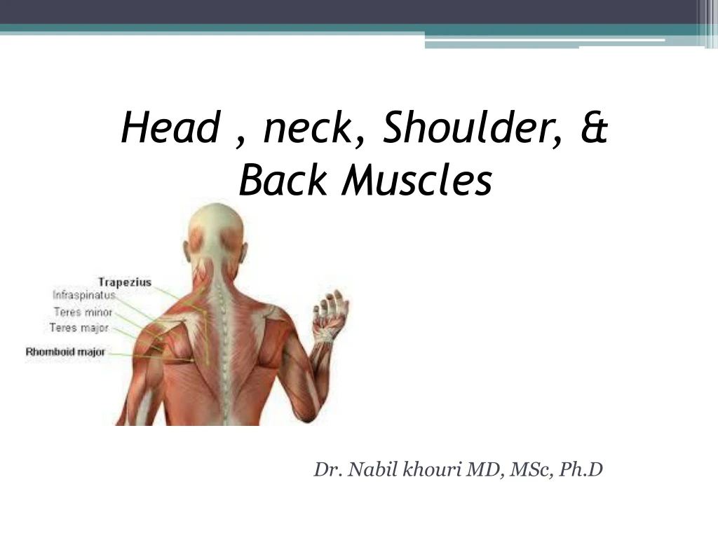
Head , neck, Shoulder, & Back Muscles Dr. Nabil khouri MD, MSc, Ph.D
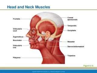
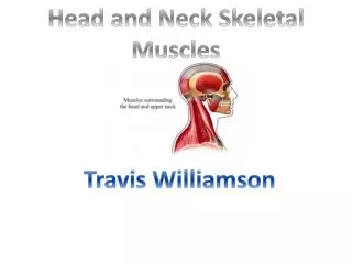
Muscles of the Head and Neck • Scalp Muscle: epicranius • frontal belly • occipital belly • gala aponeurotica • Muscles of Facial Expression: insert on skin or another muscle • Action: expression of the face • Muscles of Mastication (chewing muscles): all have insertions on the mandible • Tungue muscles • Anterior Neck Muscles • Posterior Neck Muscles
Mastication: • Jaw closure: masseter and temporalis • Side to side grinding: Lateral and medial pterygoids • Buccinator: compresses cheek Deep chewing muscles Tongue itself (instrinsic muscles): digestive tract section
Deep Muscles of mastication and deglutition Pharyngeal constrictors
Extrinsic tongue muscles “glossus” = tongue
Muscles of the Anterior Neck Suprahyoid: form floor of oral cavity, anchor tongue, elevate hyoid, move larynx superiorly during swallowing Infrahyoid: Depress hyoid and larynx during swallowing and speaking
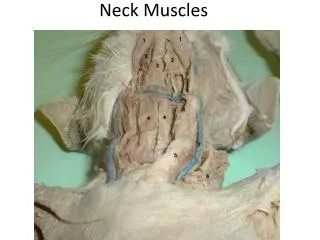
Neck muscles Posterior neck Splenius’ (capitis and cervicis) extend head Antero-lateral neck Scalenes elevate first 2 ribs
Muscles of Neck innervations • Trapezius and sternocleidomastoid (SCM) are both innervated by spinal accessory nerve (CN XI). • Suboccipitals are innervated by suboccipital nerve. • Scalenes, prevertebrals, and splenius capitis/cervicis are innervated by cervical spinal nerves. • Platysma is involved with facial expression are innervated by the facial nerve (CN VII). • Infrahyoids innervated by cervical nerves. • Suprahyoids innervated by cranial nerves.
Trapezius O: EOP, Superior Nuchal Line, Nuchal ligament, and the SP of C7 through T12 I: Upper Traps: Lateral 1/3 of clavicle & Acromion Middle Traps: Spine of scapula and acromion Lower Traps: Root of the Spine of the scapula A: Upper Traps: Elevates, upwardly rotates, and retracts the scapula Middle Traps: Retracts the scapula Lower Traps: Depresses, upwardly rotates, and retracts the scapula **Reversed muscle action: Bilaterally allows for extension of the neck. Unilaterally laterally flexes the neck to the same side and rotates to the opposite side. N: CN XI (Spinal accessory nerve) and posterior rami of C3 and C4
Splenius Capitis O: Nuchal ligament and the SP’s of C7-T4 I: Mastoid process and the occiput A: Bilateral contraction: Extension of the neck Unilateral contraction: Lateral flexion and Ipsilateral rotation of the neck N: posterior rami of the cervical spinal nerves
Splenius Cervicis • O: SP’s of T4 – T6 • I: TP’s of C1 – C3 • A: Bilateral contraction: Extension of the neck • Unilateral contraction: Lateral • flexion and Ipsilateral rotation of the neck • N: posterior rami of the cervical spinal nerves
Levator Scapulae O: TP’s of C1 – C4 I: Medial border of the scapula, from the superior angle to the root of the spine of the scapula A: Elevates retracts, and Downwardly, rotates scapula. Bilaterally allows for extension of the neck. Unilaterally laterally flexes the neck to the same side and rotates the to the same side. N: Dorsal Scapular nerve
Muscles of the Posterior Neck – Deep (Suboccipitals) • Suboccipitals are found deep to trap, SCM, splenius capitis, and semispinalis capitis. • Suboccipitals are more important as postural muscles, providing fine control of head posture, than movers. • Rectus Capitis Posterior Major: • Rectus Capitis Posterior Minor: • Obliquus Capitis Inferior: • Obliquus Capitis Superior
Rectus Capitis Posterior Major O: SP of the Axis (C2) I: Occiput (lateral aspect) A: Bilateral contraction: Extension of Head Unilateral contraction: Lateral flexion and Ipsilateral rotation of neck N: Suboccipital nerve
Rectus Capitis Posterior Minor O: Posterior tubercle of the Atlas (C1) I: Occiput A: Bilateral contraction will cause Extension of Head N: Suboccipital nerve
Obliquus Capitis Inferior O: SP of the Axis (C2) I: TP of the Atlas (C1) A: Ipsilateral Rotation of Atlas N: Suboccipital nerve
Obliquus Capitis Superior O: TP of the Atlas (C1) I: Occiput (between the superior and inferior nuchal lines) A: Bilateral Contraction: Extension of Head Unilateral Contraction: Lateral flexion of the head. N: Suboccipital nerve
Muscles Crossing the Shoulder Nine muscles cross the shoulder joint and insert into the humerus Prime movers include: Pectoralis major – arm flexion Latissimus dorsi and posterior fibers of the deltoid – arm extension Middle fibers of the deltoid – arm abduction
Deltoidmuscle • posterior fibers of the deltoid – arm extension • Middle fibers of the deltoid – arm abduction
More Muscles Crossing the Shoulder Rotator cuff muscles – supraspinatus, infraspinatus, teres minor, and subscapularis Function mainly to reinforce the capsule of the shoulder Secondarily act as synergists and fixators The coracobrachialis and teres major: Act as synergists Do not contribute to reinforcement of the shoulder joint
Sub-scapularisMuscle Origin Medial two thirds of the subscapular fossa of scapula Lower two thirds of lateral border of scapula Insertion Lesser tubercle of humerus Anterior part of joint capsule of shoulder Nerve Supply Upper subscapular nerve (C5, C6) Lower subscapular nerve (C5, C6) Actions Medial rotation of humerus Extension when shoulder fully abducted/flexed Stabilisation and protection of the shoulder joint
Trunk Movements: Deep Back Muscleshttp://cnx.org/contents/ff1ab679-4c9d-4b8f-9792-6da5fdba00d3@3 The prime mover of back extension is the erector spinae Erector spinae, or sacrospinalis, muscles consist of three columns on each side of the vertebrae – iliocostalis, longissimus, and spinalis Lateral bending of the back is accomplished by unilateral contraction of these muscles Other deep back extensors include the semispinalis muscles and the quadratus lumborum
Deep muscles of back Right side: deeper Erector spinae (extend back): Iliocostalis Longissimus Spinalis Quadratus lumborum (lateral flexion) Labeled cervicis, thoracics, lumborum depending on where they are