Download
1 / 37
460 likes | 1.18k Views
Interactions of charged particles with the patient. The depth-dose distribution - How the Bragg Peak comes about - (Thomas Bortfeld) The lateral dose distribution - Dose calculation issues - (Bernard Gottschalk). Course Outline. How the Bragg peak comes about. 1) Energy loss

E N D
Interactions of charged particles with the patient The depth-dose distribution- How the Bragg Peak comes about - (Thomas Bortfeld) The lateral dose distribution- Dose calculation issues - (Bernard Gottschalk)
How the Bragg peak comes about 1) Energy loss • collisions with atomic electrons 2) Intensity reduction • nuclear interactions W.R. Leo: Techniques for Nuclear & Particle Physics Experiments 2nd ed. Springer, 1994 T. Bortfeld: An Analytical Approximation of the Bragg Curve for Therapeutic Proton Beams, Med. Phys. 24:2024-2033, 1997
Energy loss • Protons are directly ionizing radiation (as opposed to photons) • Protons suffer some 100,000s of interactions per cm • They will eventually lose all their energy and come to rest
200 MeV, 26.0 cm 150 MeV, 15.6 cm 100 MeV, 7.6 cm 50 MeV, 2.2 cm Energy loss: Energy-range relationship, protons in water Depth 10 cm 20 cm 30 cm
Energy loss: Energy-range relationship, protons in water Convex shape à Bragg peak
Energy loss: Energy-range relationhip • General approximate relationship:R0 = aE0p • For energies below 10 MeV:p= 1.5 (Geiger’s rule) • Between 10 and 250 MeV:p= 1.8 • Bragg-Kleeman rule:a = c (Aeff)0.5/r
Energy loss: Depth dependence of the energy • Protons lose energy between z = 0 and z = R0 in the medium • At a depth z the residual range isR0 - z = aEp(z) • E(z) = a-1/p (R0 - z)1/p • This is the energy at depth z
Energy loss: Stopping power • Stopping power: • The stopping power is (within certain approximations) proportional to the dose
Energy loss: Stopping power (Dose = Stopping power)
Energy loss: Stopping power • Stopping power: • Expressed as a function of the energy:
charge of projectile electron densityof target ionization potential Energy loss: Stopping power • Bethe-Bloch equation:
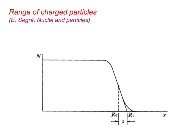
Energy loss: Range straggling • So far we used the continuously slowing down approximation (CSDA) • In reality, protons lose their energy in individual collisions with electrons • Protons with the same initial energy E0 may have slightly different ranges:“Range straggling” • Range straggling is Gaussians approx. 1% of R0
Convolution for range straggling Theoreticalw/o Straggling Range Straggling Distribution = ? *
Convolution for range straggling Theoreticalw/o Straggling Range Straggling Distribution Real Bragg Peak = * Parabolic cylinder function
Energy loss: Range straggling With consideration of range straggling
Intensity reduction: Nuclear interactions • A certain fraction of protons have nuclear interactions with the absorbing matter (tissue), mainly with 16O • Those protons are “lost” from the beam
Intensity reduction: Nuclear interactions Rule of thumb: 1% loss of intensity per cm (in water)
Intensity reduction: Nuclear interactions • Nuclear interactions lead to local and non-local dose deposition (neutrons!)
Before collision After collision Proton Proton 15O O 16 Neutron Atomic nucleus of tissue Target fragment PET isotope activation by protons • Positron Emission Tomography (PET) is potentially a unique tool for in vivo monitoring of the precision of the treatment in ion therapy • In-situ, non-invasive detection of +-activity induced by irradiation • Mainly 11C (T1/2 = 20.3 min) and 15O (T1/2 = 121.8 s) E=110 MeV Dose proportionality:A(r) ≠ D(r) 15O, 11C, ...
The Bragg curve z80=R0 T. Bortfeld, Med Phys 24:2024-2033, 1997
Protons vs. carbon ions (physical dose) Wilkens & Oelfke, IJROBP 70:262-266, 2008
Tissue inhomogeneities:A lamb chop experiment © A.M. Koehler, Harvard Cyclotron
Proton range issues:Range uncertainties due to setup Jan 08 Chen, Rosenthal, et al., IJROBP 48(3):339, 2000
Proton range issues:Range uncertainties due to setup Jan 11 Chen, Rosenthal, et al., IJROBP 48(3):339, 2000
Proton range issues:Tumor motion and shrinkage Initial Planning CTGTV 115 cc 5 weeks later GTV 39 cc S. Mori, G. Chen
Is not always what you get Beam overshoot Proton range issues:Tumor motion and shrinkage What you see in the plan… Beam stops at distal edge S. Mori, G. Chen
Proton range issues:Reasons for range uncertainties • Differences between treatment preparation and treatment delivery (~ 1 cm) • Daily setup variations • Internal organ motion • Anatomical/ physiological changes during treatment • Dose calculation errors (~ 5 mm) • Conversion of CT number to stopping power • Inhomogeneities, metallic implants • CT artifacts
Tissue inhomogeneities Goitein & Sisterson, Rad Res 74:217-230 (1978)
Tissue inhomogeneitiesBragg Peak degradation in the patient M. Urie et al., Phys Med Biol 31:1-15, 1986
Problems • Consider the proton treatment of a lung tumor (density r = 1) with a diameter of 2 cm. The tumor is surrounded by healthy lung tissue (r = 0.2). The treatment beam is designed to stop right on the edge of the tumor. After a couple of weeks the tumor shrinks down to 1.5 cm. By how much does the beam extend into the healthy lung now? • Consider a hypothetical world in which the proton energy is proportional to the proton range. How would that affect the shape of the Bragg peak?