Download
1 / 47
490 likes | 807 Views
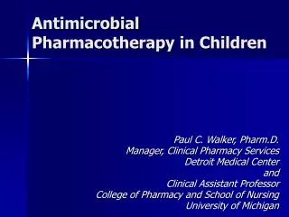
Antimicrobial Pharmacotherapy in Children. Paul C. Walker, Pharm.D. Manager, Clinical Pharmacy Services Detroit Medical Center and Clinical Assistant Professor College of Pharmacy and School of Nursing University of Michigan. Classifying Antimicrobial Agents.

E N D
Antimicrobial Pharmacotherapy in Children Paul C. Walker, Pharm.D. Manager, Clinical Pharmacy Services Detroit Medical Center and Clinical Assistant Professor College of Pharmacy and School of Nursing University of Michigan
Classifying Antimicrobial Agents • Inhibition of cell wall synthesis • Altering cell membrane permeability • Reversibly inhibiting protein synthesis • Irreversibly disrupting protein synthesis • Disruption of nucleic acid metabolism • Blocking essential metabolic events
Peptidoglycan Synthesis Peptidoglycan is composed of chains of peptidoglycan monomers (NAG-NAM-tetrapeptide). These monomers join together to form chains and the chains are then joined by cross-links between the tetrapeptides to provide strength.
Peptidoglycan Synthesis • New peptidoglycan synthesis occurs at the cell division plane by way of a collection of cell division machinery known as the divisome. • Bacterial enzymes called autolysins, located in the divisome, break both the glycosidic bonds at the point of growth along the existing peptidoglycan, as well as the peptide cross-bridges that link the rows of sugars together. • Transglycosidase enzymes then insert and link new peptidoglycan monomers into the breaks in the peptidoglycan. • Finally, transpeptidase enzymes reform the peptide cross-links between the rows and layers of peptidoglycan to make the wall strong
Structure of Bacterial Cell Walls Peptidoglycan Cross-links Comparison of the structure and composition of gram positive and gram negative bacterial cell walls
Inhibitors of Cell Wall Synthesis • Beta Lactam Antibiotics • Penicillins • Cephalosporins • Carbapenems • Monobactams • Vancomycin The beta lactam ring of penicillin
How Penicillins Inhibit Peptidoglycan Synthesis During normal bacterial growth, bacterial enzymes called autolysins put breaks in the peptidoglycan in order to allow for insertion of peptidoglycan building blocks (monomers of NAG-NAM-peptide). These monomers are then attached to the growing end of the bacterial cell wall with transglycosidase enzymes. Finally, transpeptidase enzymes join the peptide of one monomer with that of another in order to provide strength to the cell wall. Penicillins and other -lactam antibiotics bind to the transpeptidase enzyme and block the formation of the peptide cross-links. This results in a weak cell wall and osmotic lysis of the bacterium.
Natural Penicillins Penicillin G Penicillin V Aminopenicillins Ampicillin Amoxicillin Carboxypenicillins Ticarcillin Carbenicillin Penicillinase-Resistant Penicillins Cloxacillin Dicloxacillin Methicillin Nafcillin Oxacillin Ureidopenicillins Mezlocillin Piperacillin Beta Lactam Antibiotics: The Penicillins
First Generation Cephalothin Cefazolin Cephalexin Cephapirin Cefadroxil Cephradine Second Generation Cefaclor Cefoxitin Cefuroxime Cefotetan Cefpoxodime Cefprozil Cefonicid Cefmetazole Beta Lactam Antibiotics: The Cephalosporins • Third Generation • Cefotaxime • Ceftriaxone • Cefoperazone • Cefipime* • Cefmenoxime • Ceftizoxime • Ceftazidime • Cefdinir • Cefixime • Ceftibutin • This is classified as a “fourth” generation agent; it has gram negative activity similar to other third generation agents, but better gram positive coverage.
Beta Lactam Antibiotics: The Carbapenems and Monobactams • Carbapenems • Imipenem/Cilastatin • Meropenem • Ertapenem • Monobactams • Aztreonam
Side Effects and Adverse Reactions • Beta lactam Antibiotics • Hepatic dysfunction • Acute interstitial nephritis • azotemia, hematuria, proteinuria, fever, rash, eosinophilia • Neurotoxicity • Transient blood dyscrasias • Allergic or hypersensitivity reactions • Coagulopathy
Vancomycin • Indications: serious gram positive infections where -lactams are inappropriate (MRSA, MRSE, allergy, etc.) • Toxicities and Side Effects • Nephrotoxicity • Ototoxicity • Red Man Syndrome
Disrupters of Protein Synthesis • Bind to the ribosomal subunits to impair protein synthesis • Aminoglycosides • Chloramphenicol • Macrolides • Erythromycin • Clarithromycin • Azithromycin • Clindamycin
Structure of the antibiotic gentamicin C1a bound to its RNA target. Aminoglycoside antibiotics cause misreading of the genetic code. Blocks initiation of protein synthesis Blocks translation to cause premature termination Causes incorporation of incorrect amino acid The Aminoglycosides • Kanamycin • Gentamicin • Tobramycin • Amikacin • Netilmicin • Sisomycin Aminoglycosides bind to the 30s subunit to impair protein synthesis.
Agents that Bind to the 50S Ribosome • Chloramphenicol • spectrum of activity • S. pneumonia • H. influenza • Neisseria spp. • Salmonella • Bordetella • Enterobacteriaceae • some anaerobes
Macrolides Erythromycin S. pneumonia, S. pyogenes, Legionella, Chlamydia trachomatis, M. catarrhalis, H. influenza, Mycoplasma pneumonia Clarithromycin MAC Azithromycin MAC Clindamycin aerobic gram-positive bacteria anaerobes, especially B. fragilis used in combination with aminoglycosides to treat intra-abdominal and gynecologic infections Agents that Bind to the 50S Ribosome
Chloramphenicol Gray syndrome Dose-dependent bone marrow suppression Aplastic anemia, pancytopenia Macrolides GI complaints Rash Clindamycin Diarrhea Pseudomembranous colitis Rash, urticaria Hypotension Side Effects and Adverse Reactions
Disrupters of Nucleic Acid Metabolism • Metronidazole • Quinolones: • Ciprofloxacin • Levfloxacin • Moxifloxacin • Norfloxacin • Ofloxacin • Trovafloxacin • Gatifloxacin • Grepafloxacin
Disrupters of Nucleic Acid Metabolism • Metronidazole Participates in redox reactions; it is activated by a reduction of the nitro group to an anion radical. In the case of metronidazole, reduced ferredoxin appears to be the primary electron donor responsible for its reduction The anion radical is highly reactive and will form adjuncts with proteins and DNA leading to a loss of function. In particular, the reactions with DNA result in strand breakage and inhibition of replication and will lead to cell death.
Disrupters of Nucleic Acid Metabolism • Quinolones: inhibit DNA-gyrase and topoisomerase II • Ciprofloxacin • Levfloxacin • Moxifloxacin • Norfloxacin • Ofloxacin • Trovafloxacin • Gatifloxacin • Grepafloxacin
Metronidazole dizziness paresthesias peripheral neuropathy disulfiram-like reaction blood dyscrasias Quinolones headache rash, photosensitivity GI complaints arthralgias confusion liver dysfunction Side Effects and Adverse Reactions
Antimetabolites • Trimethoprim • Sulfonamides • Sulfamethoxazole • Sulfisoxazole Inhibition of folate metabolism by sulfonamides and trimethoprim
Sulfonamides Dizziness, headache Rash Blood dyscrasias Crystalluria Acute nephropathy Bilirubin displacement Side Effects and Adverse Reactions
Proper Antimicrobial Selection: Factors to Consider • Identity of infecting organism • Susceptibility of infecting organism • Host Factors
Target site modification(intracellular or extracellular; -lactams, macrolides, quinolones, glycopeptides) Decreasedpermeability(-lactams) Enzymatic degradation(intracellular or extracellular; -lactams, aminoglycosides) X Efflux(macrolides, quinolones) Bypass(TMP/SMX) Major Mechanisms of Antimicrobial Resistance
Enzyme Inactivation of Penicillins 1 = Site of action of penicillinase 2 = Site of action of amidase A = Thiazolidine ring B = -lactam ring 2 1 Structure of penicillins and interaction with beta lactamase
Betalactamase Resistance to Penicillin in N. gonorrhea
Gram Negative Organisms H. Influenza M. Catarrhalis Enterobacter Klebsiella Citrobacter Serratia Gram Positive Staphylococcus S. aureus S. epidermidis Streptococcus S. pneumoniae Vancomycin Enterococci E. faecalis E. faecium S. aureus Bacterial Resistance: What Problems are We Seeing?
Other Important Factors: MICs and MBCs Fail to Tell the Whole Story • Antimicrobial Pharmacodynamics • attempt to characterize the relationship between ANTIMICROBIAL EXPOSURE (concentration, dose, AUC) and ANTIMICROBIAL EFFECT(eg., rate, extent, and duration of antimicrobial activity)
Other Important Factors: MICs and MBCs Fail to Tell the Whole Story • Antibiotic Pharmacodynamics • Rate and Extent of Bactericidal Action • Post-antibiotic Effect • Effects of Sub-inhibitory Concentrations • Post-antibiotic Leukocyte Effect • Inoculum Effect
Classification Based on Pharmacodynamic Characteristics • Concentration-Dependent Agents • Bactericidal activity is dependent on concentration above the MIC achieved, increasing with increasing concentration • Time-Dependent Agents • Bactericidal activity is dependent on how long the concentration exceeds the MIC • Bacteriostatic Agents • Abort bacterial growth and allow host defenses to eradicate organisms
Antibiotic conc 8 7 control 6 1/4MIC 1 / log CFU per mL 5 1 MIC 4 4 MIC 16 MIC 3 64 MIC 2 1 0 1 2 3 4 5 6 Time (hours) Concentration-Dependent Killing of Pseudomonas aeruginosa with Tobramycin
9 Antibiotic conc 8 7 control 6 1/4 MIC 1 / log CFU per mL 5 1 MIC 4 4 MIC 16 MIC 3 64 MIC 2 1 0 1 2 3 4 5 6 Time (hours) NON-Concentration-Dependent Killing
Pharmacodynamic Properties by Antibiotic Class TIME dependent killing CONCENTRATION dependent killing Aminoglycosides β-lactams Fluoroquinolones GlycopeptidesAzithromycin? Metronidazole Macrolides (except Azithromycin) Chloramphenicol Rifampin Tetracyclines Clindamycin
CIDAL activity STATIC activity Bacterial REGROWTH PAE Plasma Conc Site Conc MBC MIC Pharmacodynamic Relationships between Antibiotic Concentration and Antibacterial Effect Time
Cmax Plasma Conc AUC AUC > MIC MIC T > MIC PAE Time Pharmacodynamic Relationships between Antibiotic Concentration and Antibacterial Effect
Pharmacokinetics Susceptibility MIC / MBC Serum / Tissue Concentrations Pharmacodynamics Time > MIC Peak / MIC AUC > MIC Eradication/Cure
Antibiotic Pharmacodynamics in Otitis Media: T>MIC Average percentage of time drug concentration exceeds the minimum inhibitory concentration (%T>MIC) for pediatric dosages of oral ß-lactam agents against penicillin-sensitive (black bars) and penicillin-intermediate (hatched bars) Streptococcus pneumoniae. Rodvold. Pharmacoatherapy. 2001; 21(11s) :319s-330s.
Antibiotic Pharmacodynamics: Ciprofloxacin AUC0-24:MIC and Clinical Outcomes Percentage of bacteriologic (black bars) and clinical (hatched bars) cures as a function of AUC0-24:MIC in 68 patients with gram-negative infections treated with ciprofloxacin. Note that the bacteriologic and clinical outcomes are better with AUC > 125.
Clinical Breakpoints Clinical breakpoints are supposed to indicate at which MIC the chance of eradication or even clinical success of antimicrobial treatment prevails significantly over failure, given the dosing schedule of the drug. The breakpoint thus is not only dependent on the antimicrobial activity of the drugs itself, but also on its pharmacokinetics and pharmacodynamics.
Postantibiotic effect • The period of time where there is persistent suppression of bacterial growth following exposure to an antimicrobial agent, despite removal of the antimicrobial agent.
Antibiotic 1 Antibiotic 2 Antibiotic Pharmacodynamics • MIC = minimum inhibitory concentration • MBC = minimum bactericidal concentration From: Levinson ME. Infect Dis Clin North Amer. 1995; 483-95.
Antibiotic Combinations:Rationale and Indications • Additive Effects • Synergistic Effects • Antagonistic Effects
Antibiotic Combinations:Rationale and Indications • Prevent emergence of resistance • Polymicrobial infections • Empiric therapy • Reduced drug toxicity • Synergism
Antibiotic Combinations:Disadvantages of Inappropriate Combination Therapy • Antagonism • Increased drug costs • Adverse drug reactions