Neuron Pathways: Nervous System Essentials
Explore the fascinating world of neurons, synaptic transmission, and neurotransmitters. Discover the key brain measurement methods and essential chemicals like Acetylcholine and Dopamine. Unravel the mysteries behind the central and peripheral nervous systems and dive into the brain's intricate structures like the frontal lobe. Get insights into how the brain functions and controls various bodily activities.

Neuron Pathways: Nervous System Essentials
E N D
Presentation Transcript
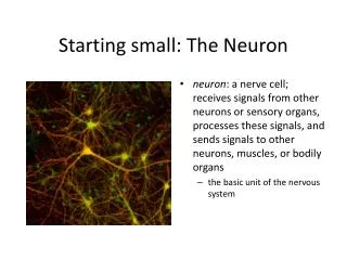
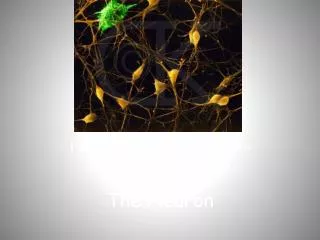
The Neuron Pathways to the Nervous System
The Neuron • 200 billion before birth • Approximately 100 billion upon birth • 90% of your cells are glia cells (in CNS) and Schwann cells (in PNS) • (fun fact: prenatal neurons develop at the rate of 250,000/minute!!)
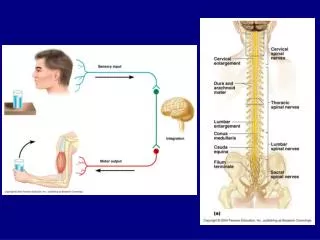
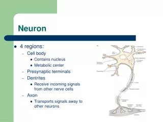
Nerve fibers are long dendrites and axons bundled together. They are called nerves in the PNS and tracts in the CNS Nerves that lead to the brain are also known as sensory, afferent or cranial nerves Nerves going from the brain are efferent or motor neurons
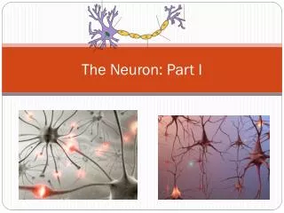
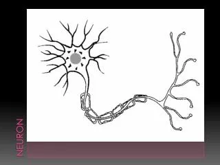
Parts: dendrites soma nucleus, nissl substance axon myelin sheath nodes of Ranvier terminal feet terminal buttons Neuron
Chemicals contained in the terminal buttons are called neurotransmitters, which will leave the terminal buttons and will synapse onto new neurons The area between neurons is known as the synaptic cleft
The all-or-none law states that when a neuron is stimulated, it must stimulated to -50 millivolts or the neuron will not fire All-or-none law
Fun fact: Approximately 99% of all information entering through the senses is immediately dropped
Ways of Measuring the Brain Electroencephalograph-EEG- Measures electrical activity of the brain Used in sleep research, detects dreams and sleep disorders
Ways of Measuring the Brain Lesioning Intentional damage to brain cells to measure the effects of the damage Lesioning has taught us a lot about the functions of the brain
CT-Computerized Tomography X-Ray of the brain Horizontal slices-tells us about damage Least expensive – widely used Ways to Measure the Brain
Positron Emission Tomography (PET) Radioactive tracer Measure blood flow and metabolic activity in brain Color coded map Ways to Measure the Brain
Magnetic Resonance Imaging (MRI) Magnetic fields and radio waves 3-D image of the brain Very expensive Ways to Measure the Brain
Ways of Measuring the Brain Electrical Stimulation of the Brain (ESB) Sends a weak electrical current to brain Stimulates areas of the brain to determine what effect the stimulation has Discovered by Penfield, done during brain surgery
Acetylcholine-Ach Involved in voluntary movements Contributes to attention, arousal and memory Lack of Ach – implicated in Alzheimer’s Low levels of Ach – cells die
Acetylcholine Hippocampus of the brain Overabundance of Ach-stimulation of memory and arousal Curare-a poison that blocks the transmission of Ach, which will lead to intense spasms of the muscles, including heart – can paralyze people
Controls voluntary muscles A monoamine Lack of – Parkinson’s (muscle tremors, rigidity, loss of control of muscle movements) Too much – Schizophrenia (irrational thoughts, hallucinations, break from reality) Dopamine-DA
Dopamine Tardive Dyskenesia-a disease which can cause Parkinson’s-like tremors. This occurs when a person takes meds like L-dopa for Schizophrenia Plays a major role in addiction as it is similar to adrenaline and controls the ability to feel pleasure and pain
Dopamine Dopamine agonists bind to dopamine receptors to stimulate those receptors (for Parkinson’s) Dopamine antagonists are drugs that bind but don’t stimulate dopamine receptors. Instead, they prevent or reverse the actions of dopamine by not allowing it to attach to receptors (for Schizophrenics)
Highly implicated in Depression, hence your SSRI’s (antidepressants) Low levels of SE seemed to be linked to Depression and OCD Involved in wakefulness and sleeping, memory, control of appetite A Monoamine Serotonin-SE
Norepinephrine-NE Low levels of NE have been associated with depression Cocaine and amphetamines may stimulate the activity of this neurotransmitter (may cause hallucinations) A monoamine
Synthesized by the adrenal gland Activates the sympathetic nervous system and puts the body into fight-or-flight Epinephrine
Resemble opiates “runner’s high” Pain blockers Gate control theory Endorphins
Low levels – anxiety Antianxiety drugs increase levels of GABA GABA
Central Nervous System – Consists of the: Brain Spinal cord Back of the retina Central and Peripheral Nervous Systems
Contains: Somatic Nervous System Voluntary skeletal muscles Autonomic Nervous System Sympathetic NS Parasympathetic NS Sympathetic NS Peripheral Nervous System
Parasympathetic Rest and digest response Brings the system back to homeostasis Peripheral Nervous System
Did you know that the brain weighs only about 1 pound at birth? By one year of age, the brain will double in size and be almost 90% of its adult size The Brain
FRONTAL LOBE Located in front of the central sulcus. Concerned with reasoning, planning, parts of speech and movement (motor cortex), emotions, and problem-solving. Lobes of the Brain
Frontal Lobe- On the left side is Broca’s area – damage to this area may result in Broca’s aphasia – inability to speak Lobes of the Brain
After Gage had an accident in which a metal pipe went through his brain, it was thought that he would not survive. However, he did – with interesting consequences Phineas Gage
PARIETAL LOBE Located behind the central sulcus. Concerned with perception of stimuli related to touch, pressure, temperature and pain. Lobes of the brain
The Primary Somatosensory Cortex Neurons in the primary somatosensory are activated when the skin is touched. However, the body is NOT represented in the cortex in proportion to the amount of skin.
A map of the human somatosensory cortex was drawn by Dr. Wilder Penfield in the 1950's. By observing the location on the brain that caused patients to feel sensations on different parts of their bodies, Dr. Penfield was able to draw a map of the brain
TEMPORAL LOBE Located below the lateral fissure. Concerned with perception and recognition of auditory stimuli (hearing) and memory (hippocampus). Lobes of the Brain
Temporal Lobe – Behind the temporal lobe is the uncus – memory for smell Left side of temporal lobe is Wernicke’s area-damage affects comprehension of speech Lobes of the Brain
OCCIPITAL LOBE Located at the back of the brain, behind the parietal lobe and temporal lobe. Concerned with many aspects of vision. Lobes of the Brain
. The Lobes of the Brain
Brain Weight(gm) Species 6,000 Elephant 1,300-1,400 Adult Human 97 Rhesus Monkey 72 Dog 30 Cat 10 Rabbit 2.2 Owl The Brain
Cerebral cortex functions: Thought Voluntary movement Language Reasoning Perception Parts of the Brain
Cerebellum Functions: Movement-i.e., tracking a target or playing an instrument Balance Posture Looks like a mini-brain The Hindbrain
Cerebellum • Two peach-size mounds of folded tissue at the base of the brain form the cerebellum. • Damage to this area leads to motor or movement difficulties. • Some scientists have discovered cognitive problems as well.
Medulla Control of: Blood pressure Heart rate Breathing a.k.a. the medulla oblongata The Hindbrain