Download
1 / 20
200 likes | 304 Views
Explore the complexity of protein structures, folding mechanisms, reversible interactions, and protein-protein interactions. Learn about the principles of reversible interactions, quantification of protein-ligand interactions, and a case study on oxygen binding in myoglobin and hemoglobin.

E N D
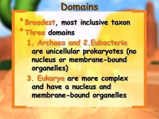
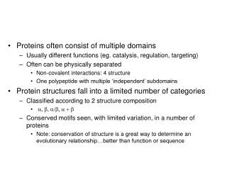
Proteins often consist of multiple domains • Usually different functions (eg. catalysis, regulation, targeting) • Often can be physically separated • Non-covalent interactions: 4 structure • One polypeptide with multiple ‘independent’ subdomains • Protein structures fall into a limited number of categories • Classified according to 2 structure composition • a, b, a/b, a + b • Conserved motifs seen, with limited variation, in a number of proteins • Note: conservation of structure is a great way to determine an evolutionary relationship…better than function or sequence
Protein folding is complex • How does a protein “know” how to fold? • Completely due to amino acids (some proteins may need assistance from molecular “chaperones”) • Studying protein folding • Often through denaturation/renaturation curves: how stable is a protein? How quickly does it (un)fold? • Several imperfect models
Reversible binding involving proteins • Interactions between proteins • Protein/DNA • Protein/small molecule ligand
Reversible binding involving proteins (Ch. 5) • Interactions between proteins • Different from 4° structure • Lower affinity (in general) • Reversible • Potential for numerous partners
Protein-protein interaction 4° Structure Hemoglobin Four ‘separate’ polypeptide chains One ‘protein’ Function as a whole Antibody (green)/Antigen (red) Two different proteins Found apart
Reversible binding • asdf • Protein vs. “small” molecule • Protein acts as a carrier for the molecule • Hemoglobin/O2 • Metallochaperones • Enzymes • Catalyze a reaction involving the substrate • Protein-DNA interactions
Principles of reversible interactions • Affinity of protein for ligand is very specific • eg. high affinity for Mg2+, low affinity for Zn2+ • eg. fumarase: distinguishes stereoisomers of tartaric acid • Ligand binding site is usually complementary to the ligand BUT ligand binding can cause drastic conformational changes • Induced fit • Conformational changes result in tighter binding but strain both protein and ligand
C C C C cAMP binding results in conformational change: regulatory subunits no longer bind catalytic: ACTIVE PKA INACTIVE PKA
Principles of reversible interactions • Enzymes • Ligands = substrate and product • Induced fit stress can drive catalysis
Quantification of protein-ligand interactions (non-catalytic) P + L ↔ PL Reversible: represent as equilibrium [PL] [P][L] Association constant (don’t confuse with Ka/pKa) Ka = High Ka: [complex] is relatively high ie. protein has a high affinity for the ligand [PL] [P] Amount of complex depends on concentration of free ligand as well as the affinity (Ka) Ka *([L]) =
Quantification of protein-ligand interactions • Work with dissociation constants PL ↔ P + L Equilibrium equation describing dissociation Kd = [P][L] Note that Kd = 1/Ka [PL]
Quantification of protein-ligand interactions • Assume [L] >> [P] • Few proteins (binding sites), lots of the ligand • ie. conc. of free ligand doesn’t change (much) even if all ligand-binding sites are filled • Fraction of ligand binding sites filled (q) q = [L] [L] + Kd
q = [L] [L] + Kd When [L] = Kd, q = 0.5 **When [L] = Kd (note: no matter what [P] is (remember assumption, though)), half of the binding sites will be filled Lower Kd: need less ligand to fill binding sites Lower Kd corresponds to higher affinity/stronger binding
% of sites filled vs. [L] Units of Kd: concentration (M, mM, mM, etc)
Protein x with three different ligands Max binding (q = 1.0, all binding sites filled/saturated) 1 Fraction of binding sites occupied q 50% of saturation 0.5 0 Kd2 Kd1 Kd3
Case study: oxygen binding in myoglobin and hemoglobin • Oxygen is poorly soluble in water (blood) • Iron (Fe2+)/O2 complex is soluble • But free iron is toxic • Use proteins containing an iron cofactor • Myoglobin • Hemoglobin
Iron is part of a heme prosthetic group: permanent association with protein
Iron has six coordination sites Four bind heme nitrogens One binds protein histidine “proximal” histidine One can bind O2
Structure of myoglobin • Extremely compact • ~75% a helix (no b structure) • Eight helical segments • Four terminate in proline • Interior: hydrophobic except for two histidines