Download
1 / 110
1.11k likes | 1.14k Views
Discover the definition and classification of vasculitis, from large vessel to small vessel and various types of affected organs. Learn about the common clinical manifestations and mimics of vasculitis. Gain insights into the etiological and segmental classifications.

E N D
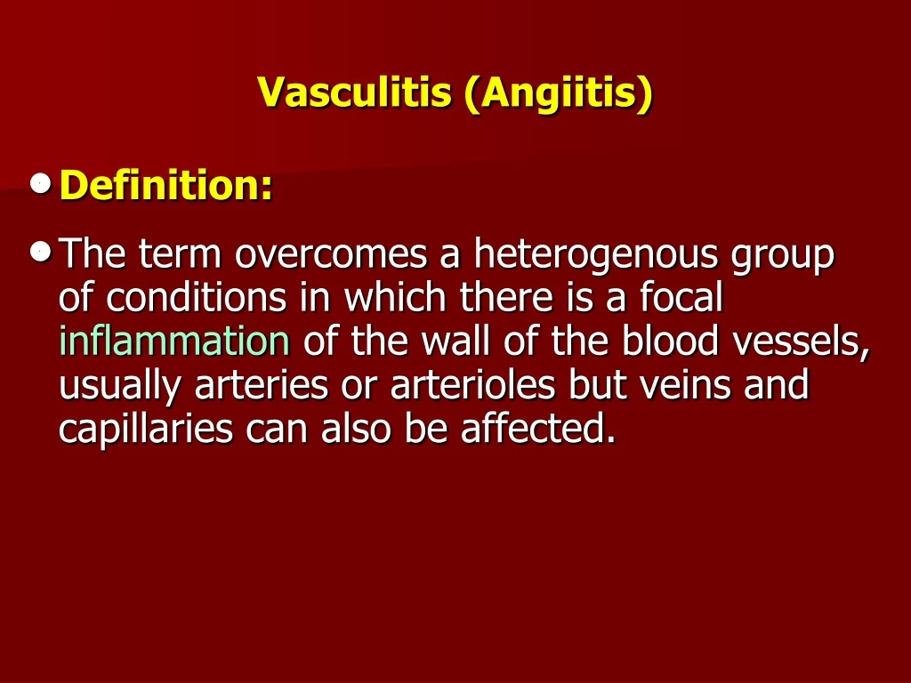
Vasculitis (Angiitis) • Definition: • The term overcomes a heterogenous group of conditions in which there is a focal inflammationof the wall of the blood vessels, usually arteries or arterioles but veins and capillaries can also be affected.
Artery: WBC inflammation in wall Normal Artery
General consideration: • Vasculitis or vasculitides are characterized by inflammation and necrosis of blood vessels. • Vessels of any size and of any organ may be affected. • This is reflected in the wide variety of signs and symptoms that characterize vasculitis. • Clinically vasculitides range from a benign, regionally restricted process to a life-threatening systemic disorder. • Thus, they can mimic or be mimicked by a variety of diseases.
Therefore, a systematic approach to correct diagnosis is necessary. • In severe arteritis there is necrosis of the arterial wall, which may result in occlusive thrombosis, rupture or aneurysm formation. • The healing phase often causes severe permanent narrowing of the lumen, resulting either from organization of thrombus or from endarteritis obliterans, resulting in chronic ischemia.
Classification of vasculitis • Based on the site of the affected vessels, vasculitis is classified into: • Skin only e.g. Cutanousvasculitis • Internal organ only e.g. Isolated angiitis of CNS. • Both e.g. Systemic vasculitis
Based on the size of the affected vessels, vasculitis is classified into: • Large vessel Vasculitis: • Giant cell arteritis (GCA) • Temporal arteritis (TA) • Takayasu’sarteritis (TD)
Medium vessel Vasculitis • Classic polyarteritisnodosa (PAN) • Kawasaki’s disease (KD) • Thromboangitisobliterans (TAO) • Small vessel Vasculitis • Hypersensitivity vasculitis • HenochSchonleinpurpura (HSP) • Churg Strauss syndrome (CSS) • Wegener granulomatosis(WG) • Microscopic polyangiitis (MPA)
Large vessel vasculitis: usually involves aorta and its larger branches to extremities and head and neck. • Medium sized vessel vasculitis: involves arteries of the viscera and their branches. • Small vessel vasculitis (most are due to immune complex deposition; type III hypersensitivity reaction): involves arterioles, venules and capillaries.
Based on the size of the lesion, vasculitis is classified into: • Focal (causing aneurysm) • Segmental (causing stenosis or occlusion)
Etiological classification of small vessel vasculitis involving the capillaries: • ANCA associated vasculitis. • Immune complex mediated vasculitis. • Paraneoplastic small vessel vasculitis. • Inflammatory bowel disease associated vasculitis.
ANCA associated: • Microscopic polyangiitis (MPA) • Wegener granulomatosis (WG) • Churg Strauss syndrome (CSS) • Drug-induced vasculitis
Immune complex mediated: • Henoch-Schönlein purpura (HSP) • Essential cryoglobulinemicvasculitis (ECV) • Systemic lupus vasculitis • Rheumatoid vasculitis • Sjogren’s syndrome vasculitis • Hypocomplimentemicurticarialvasculitis • Behcet’s disease • Goodpasture’s syndrome • Serum sickness vasculitis • Infection induced vasculitis • Cutaneousleucocytoclastisvasculitis • Drug-induced vasculitis
Paraneoplastic small vessel vasculitis: • Lymphoproliferative, myeloproliferative neoplasm induced vasculitis • Carcinoma-induced vasculitis • Inflammatory bowel disease associated vasculitis.
Mimicks of vasculitis • Bacterial endocarditis • Antiphospholipid antibody syndrome • Viral infections like HIV, CMV, HBV, HCV • Cholesterol emboli syndrome • Vasoconstrictor drugs (ergot poisoning) • Hypertensive arteriopathy • Thoracic outlet syndrome • Disseminated intravascular coagulation (DIC) • Sepsis
Sarcoidosis • Thrombotic thrombocytopenic purpura (TTP) • Lymphoma, hairy cell leukemia
Common Clinical Manifestations • Systemic or general features: Fever, sweats, malaise. weight loss. • Skin:Palpable purpura. • Neurologic: Mononeuritis multiplex. • Brain: Stroke. • Musculoskeletal:Arthralgia/arthritis, muscle pain/claudication. • Respiratory:Sinusitis/epistaxis, pulmonary infiltrates.
Gastrointestinal: Abdominal pain, bloody stool. • Renal: Glomerulonephritis, hypertension. • Cardiovascular system:Myocardial infarction, coronary aneurysms.
Clinical manifestations of vasculitis depends on the size and extent of the vessel involvement: • Large vessel vasculitis: • Presents with loss of pulse or stroke • Medium vessel vasculitis • Presents with infarction or aneurysm • Small vessel vasculitis • Presents with palpable purpura
Loss of pulse: in the upper extremity due to involvement of subclavian artery. • Vasculitis→ narrowing of lumen → decreased blood flow → loss of pulse. • Stroke: due to involvement of internal carotid artery. • Infarction and aneurysms: due to involvement of medium size arteries like coronary, renal, popliteal arteries.
Patients may present with myocardial infarction (coronary), renal infarction (renal artery) and ischemia of lower limb (popliteal artery). • Inflammation in the vessel wall may damage it and lead to formation of aneurysm e.g. coronary artery aneurysm seen in Kawasaki disease. • Purpura: are small areas of hemorrhage in the skin. • They are larger than petechiae.
Patients may present with myocardial infarction (coronary), renal infarction (renal artery) and ischemia of lower limb (popliteal artery). • Inflammation in the vessel wall may damage it and lead to formation of aneurysm e.g. coronary artery aneurysm seen in Kawasaki disease. • Purpura: are small areas of hemorrhage in the skin. • They are larger than petechiae.
Purpura could be seen in different clinical settings e.g., thrombocytopenia, vessel wall instability (vitamin C deficiency) and small vessel vasculitis. • Purpura due to thrombocytopenia or vessel instability is not palpable, because acute inflammation is not involved. • Small vessel vasculitis causes palpable purpura because the vessel wall is damaged and blood extravastes plus the vessel wall is inflamed “tumor of inflammation”.
Etiology of Vasculitis • In the vast majority of cases of vasculitis the etiology is unknown. • In vasculitis of SLE, rheumatoid artheritis, and serum sickness immune complex deposition was found. • In small number of cases of PAN (10%) the lesions are due to deposition of complexes containing hepatitis B surface antigens.
Other viral antigens or streptococcal antigen are implicated in other cases. • Occasionally vasculitis develops following administration of some drugs. • In these cases the drugs act as an immunogen resulting in immune complex deposition or there may be a hypersensitivity reaction following binding of the drug to endothelial cells.
Circulating anti-endothelial cell antibodies are present in some cases of vasculitis implying type II hypersensitivity reaction. • In graft rejection antibodies to major histocompatiblity complex (MHC) antigens bind to endothelium and are implicated in the vascular damage occurs. • Diagnosis of vasculitis is established on the basis of clinical and histological features, the distribution of lesions, and the presence of other diseases.
Histological features show considerable variation, but essentially comprise inflammation of the vessel wall which may be predominantly acute, a mixture of acute and chronic, or may be granulomatous. • Thus, neither satisfactory etiological nor morphological classification exists. • There is often good clinical response to immunosuppressive or cytotoxic drugs and early treatment is indicated.
Pathogenesis of Vasculitis: • Vasculitic syndromes are immune-mediated diseases. • Most are idiopathic. • Pathogenesis of vasculitis involves two processes, namely: • Inflammation of vessel wall • Ischemia of the tissues • Based on this, vasculitis may be primary or secondary.
Mechanism of vessel wall involvement: • Immunologic hypersensitivity reactions: • Type I:Allergic angiitis with eosinophilia and raised IgE levels in blood and in the tissues e.g. in Churg Strauss syndrome (CSS). • Type II: Antibody mediated (pauci-immune type) vasculitis which includes ANCA associated vasculitis (AAV) like Wegner’s granulomatosis (WG), CSS and microscopic polyangiitis (MPA).
Type III: Immune complex mediated with immune complex deposition in the tissues or formation in situ. • This is associated with low complement levels, e.g. Henoch-Schonleinpurpura (HSP), polyarteritisnodosa (PAN). • Type IV:T cell mediated hypersentivitiy characterized by an inflammatory infiltrate induced by Th1 cells, e.g. Giant cell arteritis (GCA), and Takayasu’s arthritis (TA).
Laboratory testing in vasculitis • Antineutrophilcytoplasmic antibodies (ANCA) • Erythrocyte sedimentation rate (ESR) • A high ESR in conjunction with specific findings such as tender temporal artery can be highly suggestive of giant cell arteritis.
Antineutrophilcytoplasmic antibodies (ANCAs) • ANCA are seen in some types of vasculitis especially small vessel vasculitis. • They are of key importance for classification of small vessel vasculitis. • They are circulating antibodies reactive with neutrophilcytoplasmic antigens; ANCA. • ANCAs activate neutrophils→ release of enzymes and free radicals resulting in vessel damage.
ANCA titers correlate with the disease activity. • ANCA is detected by immunofluorescence. • Estimation of ANCA in patients with clinical features suggestive of WG/CSS/MPA is recommended. • This may postpone the immediate need of invasive lungs/kidney biopsy and help in early diagnosis. • This test is neither absolutely sensitive nor specific, hence confirmation of the diagnosis requires biopsy from the involved organ.
Two types of ANCAs • Cytoplasmic (c-ANCAs): • Antibodies directed against proteinase 3 in cytoplasmic granules. • Cytoplasmic staining pattern. • Example: Wegener’s granulomatosis. • Perinuclear (p-ANCAs): • Antibodies directed against myeloperoxidase. • Perinuclear pattern of staining. • Example: Churg-Strauss syndrome, PAN.
ANCAs are not 100% sensitive nor specific. • ANCAs can be found in other conditions apart from vasculitis.
Role of ANCA C-ANCA is reasonably specific for WG (75-80%) and MPA (25-30%). It is detected in only 10-15% cases of CSS. P-ANCAis less specific, detected in MPA (50-60%), CSS (55-60%), WG (10-15%) and some other conditions like drug-induced vasculitis, rheumatoid arthritis (30-70%), SLE (20-30%), ulcerative colitis (50-70%), Crohn's disease (20-40%) and different hepatic disorders.
If C-ANCA and P-ANCA coexist in a patient, drug-induced vasculitis should be suspected. • Hence the role of ANCA is in screening for these disorders and should be asked for, only when highly suggestive clinical features like pulmonary hemorrhage, recurrent sinusitis orbital mass or glomerulonephritis are present. • Estimation of ANCA level is also helpful in disease monitoring.
ANCA is almost always negative in Takayasu's disease, temporal arteritis and Kawasaki disease. • Serology for HIV, hepatitis B and C viruses,immunological tests like rheumatoid factor (RA factor), antinuclear antibody (ANA) and anti-DNA are helpful screening tests for associated disorders • Endoscopy may be of diagnostic help in cases of HSP with gastrointestinal symptoms as presenting features.
Coin-like petechiae, hemorrhagic erosions, skip hyperemia and ecchymosis are the lesions suggestive of skin vasculitis. • In patients with palpable purpura and poorly localized colicky abdominal pain, CT scan/ ultrasonography may be helpful to assess the extent of GIT involvement. • The hallmark findings in CT scan are focal areas of bowel thickening, mesenteric edema, nonspecific lymphadenopathy and vascular engorgement.
Giant cell (temporal) arteritis • Is the most common type of vasculitis. • It is more common in female than in male. • The age of onset of giant cell arteritis is usually 70 years. It is rare before 50 years. • Therefore the age at onset helps to differentiate this condition from other vasculitis that may involve similar vessels such as Takayasuarteritis which occurs in much younger people.
Vessel involvement: • Typically involves medium and large arteries,temporal artery and extra-cranial branches of external carotid. • Involvement of ophthalmic branch of external carotid blindness. • Etiopathogenesis: • Type IV hypersensitivity mediated reaction causing granulomatous inflammation. • Affected vessel are cordlike and show nodular thickening.
Grossly: • Examination of the temporal artery of a patient with giant-cell arteritis shows a thickened, nodular, and tender segment of a vessel on the surface of head. • Giant cell arteritis: may rarely involve the aortic arch → giant cell aortitis.
Microscopy: • Focal granulomatous inflammation of temporal artery. • Giant cell arteritis involves an abnormal inflammation and fragmention of internal elastic lamina in the walls of some arteries, commonly the temporal artery and other branches of the external carotid artery. • The elastic tissue is not easily degraded and stimulates the formation of multinucleated giant cells as part of the granulomatous chronic inflammatory process.
Temporal (giant cell) arteritis Giant cell
Giant cell (temporal) arteritis • Clinical features: • Fever, fatigue, weight loss. • Temporal unilateral headache, along the course of temporal artery. • Painful, palpably enlarged and tender temporal artery. • Generalized muscular aching and joint stiffness espcially in the shoulders and hip.
Temporary/permanent blindness due to involvement of ophthalmic artery. Corticosteroids are indicated to prevent blindness. • Jaw claudication. • Investigations: • ESR: screening test of choice is markedly elevated.
Temporal artery biopsy: definitive diagnosis; it is not positive in all cases (positive in only 60% cases) as the inflammatory process in not continuous but skips areas are present. • Therfore if biopsy is obtained from the part of the artery which is not involved, positive findings would then be missing.