Download
1 / 32
320 likes | 392 Views
Learn about the functions of the pancreas, enzyme secretion, clinical significance of amylase and lipase, and bone metabolism. Understand how calcium levels impact bone function and the role of hormones like PTH and vitamin D.

E N D
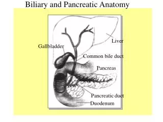
Pancreas Pancreas is a large gland Composed of both exocrine and endocrine functions 1. Endocrine functions(hormone releasing): Consist of islet of Langerhans Secrete 4 hormones (insulin, glucagon, gastrin and somatostatin) 2. Exocrine functions (enzyme secreting): Rich in digestive enzyme(amylase and lipase)produced by acinar cells
Pancreas functions Break down food using digestive enzymes of pancreas Secretes hormones that affect the level of sugar in blood Produces chemicals that neutralize stomach acid (NaHCO3)
Amylase • Amylase is an enzyme found primarily in the pancreas and salivary glands. • Its function is to assist in the digestion of complex carbohydrates into simple sugars. • Measurement of serum amylase is often performed to differentiate: • abdominal pain due to acute pancreatitis • from other causes of abdominal pain that may require surgical treatment.
Isoenzymes • Two isoenzymes are found in normal serum; • P isoamylase … derived from pancreas • S isoamylase … derived from salivary glands • They are identified using electrophoresis chromatography or isoelectric focusing.
The serum amylase begins to rise 3 to 6 hours after the onset of acute pancreatitis and peaks in approximately 24 hours. The values return to normal within 5 days after onset. For confirmation of an acute pancreatitis measurement of lipase should be additionally performed. The amylase present in the blood is eliminated through the kidney and excreted into the urine. Therefore, elevation of serum activity is reflected in a rise of urinary amylase activity.
Clinical Significance • Increased • Acute pancreatitis • Alcoholism • Hyperlipidemia • Inflammation of salivary glands • Decreased • Cystic fibrosis
Principle Enzymatic photometric test, in which the substrate EPS-G7 is cleaved by α -amylases into various fragments. These are further hydrolyzed in a second step by α -glucosidase producing glucose and p-nitrophenol. The increase in absorbance represents the total (pancreatic and salivary) amylase activity in the sample.
Principle (PNP = p-Nitrophenol, G =Glucose) 4,6-ethylidene (G7)- ρ -nitrophenyl(G1)- α,D-maltoheptaoside (EPS-G7)
Lipase (LPS) • Lipase is an enzyme that hydrolyzes the ester linkage of fats to produce alcohols and fatty acids • LPS catalyses the partial hydrolysis of dietary triglycerides in the intestine • Pancreatic LPS specific for fatty acid residues at position 1 and 3 of triglyceride • Accelerated by presence of bile salts & colipase
Lipase (LPS) • Primarily found in pancreas • Some from stomach and small intestine
Diagnostic Significance • Used in diagnosis of acute pancreatitis • Similar pattern to AMS but lasts longer • LPS elevations persist for approximately 5 days in acute pancreatitis, whereas AMS elevations persist for only 2 to3 days • May be elevated in other abdominal conditions • Normal in salivary gland conditions (AMS↑) • LPS levels are useful in differentiating serum AMS elevation as a result of pancreatic versus salivary involvement
Assay for enzyme activity • Turbidimetric methods • Fats in solution = cloudy • LPS hydrolysis causes clearing of solution • Colorimetric • Coupled reactions with peroxidase or glycerolkinase
Bone Bone is a rigid organ that constitutes part of the vertebrate skeleton.
Bone profile test Calcium Magnesium ALP
Calcium • Calcium is required for cell function overall and for bone metabolism (Approximately 99% of body calcium is found in bones). • Calcium (Ca2+) is present in the blood in two forms: • Approximately 45% is present in a free state, • and the other 55% is bound to plasma protein, primarily to albumin.
Too little calcium gets you either a loss of tissue function or soft bones (osteoporosis) and tetani. Changes in calcium are used to assess bone function. Higher blood levels usually mean lower bone levels. Usually performed in conjunction with Phosphorous determinations. Three hormones, PTH, vitamin D, and calcitonin, are known to regulate serum Ca2+ by altering their secretion rate in response to changes in ionized Ca2+.
PRINCIPLE OF THE METHOD The measurement of calcium in the sample is based on formation of color complex between calcium and o-cresolphtalein in alkaline medium: Ca++ + o-Cresolphtalein OH Colored complex O-Cresolphthalein Complex one gives violet color in alkaline medium.The intensity of the colour formed is proportional to the calcium concentration in the sample
Specimen • Serum or heparinized plasma or 24 Hrs urine • Separation from cells as rapidly as possible to avoid the uptake of calcium by erythrocytes. • Venous stasis (prolonged tourniquet application) and forearm exercise may alter calcium value due to a decrease in pH caused by localized production of lactic acid. • Exposing the sample to air will cause an increase in pH due to the loss of CO2 which will decrease ionized calcium.
HYPOCALCEMIA Vitamin D deficiency Hypoparathyroidism Alkalosis (Alkalemia) HYPERCALCEMIA Hyperparathyroidism Malignancy of bone Thyrotoxicosis Vitamin D intoxication Idiopathic
Phosphorus Phosphorus is an essential mineral for tissue bone formation and is required by every cell in the body for normal function. Approximately 85% of the body phosphorus is found in bone and in teeth. Low levels of phosphorus, can be caused by hypervitaminosis, primary hyperparathyroidism, renal tubular disorders, antacids or malabsortion. High levels of phosphorus can be caused by diet, bone metastases, liver disease, alcohol ingestion, diarrhea and vomiting Clinical diagnosis should not be made on a single test result; it should integrate clinical and other laboratory data.
Regulation • Phosphate in blood may be: • absorbed in the intestine from dietary sources, • released from cells into blood, • and lost from bone. • In healthy individuals, all these processes are relatively constant and easily regulated by: • renal excretion • or reabsorption of phosphate.
Regulation • Renal regulation is affected by factors such as: • Vitamin D, • increases both phosphate absorption in the intestine and phosphate reabsorption in the kidney • PTH, • lowers blood concentrations by increasing renal excretion.
PRINCIPLE OF THE METHOD Inorganic phosphorus reacts with molybdic acid forming a phosphomolybdic complex. Its subsequent reduction in alkaline medium originates a blue molybdenum colour. The intensity of the color formed is proportional to the inorganic phosphorus concentration in the sample
ALP Alkaline phosphatase is an enzyme present in almost all part of the organism, being particularly high in bone, liver, placenta, intestine and kidney. Both increases and decreases of plasma ALP are of importance clinically. Causes of increased plasma ALP: Paget's disease of bone, obstructive liver disease, hepatitis, hepatotoxicity caused by drugs or osteomalacia. Causes of decreased plasma ALP: Cretinism and vitamin C deficiency. .
PRINCIPLE OF THE METHOD Alkaline phosphatase (ALP) catalyses the hydrolysis of p-nitrophenyl phosphate at pH 10.4, liberating p-nitrophenol and phosphate, according to the following reaction: p-Nitrophenylphosphate+Mg++ + H20 p-Nitrophenol + Phosphate The rate of p-Nitrophenol formation, measured photometrically, is proportional to the catalytic concentration of alkaline phosphatase present in the sample
Procedure: Pipette into disposable or well clean cuvettes:
Reference range: Calculation: Activity = Abs/min * 2764