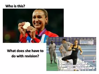
Who is this?
Who is this?. What Happened?. How much rain?. Medial Collateral Ligament. MCL. The medial collateral ligament (MCL) is one of four ligaments that are critical to the stability of the knee joint.


Who is this?
E N D
Presentation Transcript
MCL • The medial collateral ligament (MCL) is one of four ligaments that are critical to the stability of the knee joint. • A ligament is made of tough fibrous material and functions to control excessive motion by limiting joint mobility. • The MCL resists widening of the inside of the joint, or prevents "opening-up" of the knee.
Anatomy -Three layers • Superficial MCL – primary static stabilizer (under the satorial fascia) – valgus and ER • Deep MCL – middle third of medial capsule • Posterior Oblique Ligament – 3 attachments functions with semimembranosus
Dynamic stabilizers of medial knee • Semimembranous complex • Quadriceps • Pesanserine
MCL Injuries • Because the MCL resists widening of the inside of the knee joint, the ligament is usually injured when the outside of the knee joint is struck. • This force causes the outside of the knee to buckle, and the inside to widen. • When the MCL is stretched too far, it is susceptible to tearing and injury. • An injury to the MCL may occur as an isolated injury, or it may be part of a complex injury to the knee. • Other ligaments, most commonly the ACL, or the meniscus, may be torn along with a MCL injury.
Symptoms of MCL Tears • The most common symptom following an MCL injury is pain directly over the ligament. • Swelling over the torn ligament may appear, and bruising and generalized joint swelling are common 1 to 2 days after the injury. • In more severe injuries, patients may complain that the knee feels unstable, or feel as though their knee may 'give out' or buckle. • Symptoms of a MCL injury tend to correlate with the extent of the injury. MCL injuries are usually graded on a scale of I to III.
Symptoms • 67% of patients with complete tear could walk into the office unaided • Pain was worse with incomplete rather than complete
X-rays • Anteroposterior • Lateral • Merchant view
Grade I MCL Tear • This is an incomplete tear of the MCL. • The tendon is still in continuity, and the symptoms are usually minimal. • Patients usually complain of pain with pressure on the MCL, and may be able to return to their sport very quickly. • Most athletes miss 1-2 weeks of play.
Grade II MCL Tear • considered incomplete tears of the MCL. • These patients may complain of instability when attempting to cut or pivot. • The pain and swelling is more significant, and usually a period of 3-4 weeks of rest is necessary.
Grade III MCL Tear • A grade III injury is a complete tear of the MCL. • Patients have significant pain and swelling, and often have difficulty bending the knee. • Instability, or giving out, is a common finding with grade III MCL tears. • A knee brace or a knee immobilizer is usually needed for comfort, and healing may take 6 weeks or longer.
MCL injuries • Isolated • Combined with other injuries
Knee in 30 degrees of flexion compare to other knee – degree of opening and the end feel
Surgery for MCL Tears: • Surgery for MCL tears is controversial. • There are many studies that document successful nonsurgical treatment in nearly all types of MCL injuries. • patients who complain of persistent knee instability,, surgery is reasonable. • Some surgeons advocate surgical treatment of grade III MCL tears in elite athletes or in those athletes with multiple ligament injuries in the knee
Rehabilitation • Early protected ROM • Strengthening • Laxity of knee in extension – red flag
Treatment • Treatment of an MCL tear depends on the severity of the injury. • Treatment always begins with allowing the pain to subside, beginning work on mobility, followed by strengthening the knee to return to sports and activities. • Bracing can often be useful for treatment of MCL injuries. • Fortunately, surgery is not necessary for the majority
Rehabilitation Protocol • MCL injuries who require an early return to high level activity following injury. • Goals of rehabilitation are to: Control joint pain, swelling Regain normal knee range of motion Regain a normal gait pattern Regain normal lower extremity strength Regain normal proprioception, balance, and coordination • The physical therapy is to begin as soon as possible after the injury.
Phase 1: Week 1-2Range of Motion: • Passive ROM, No limits • Aggressive Patella mobility • Ankle pumps • Gastroc-soleus stretches • Wall slides • Heel slides
Strength: • Quad sets x 10 minutes • SLR (flex, abd, add) • Multi-hip machine (flex, abd, add) • Mini squats (0-45 °) • Multi-angle isometrics (90-60 °) (No tension on MCL) • When working adductors stress point should be superior to knee • Calf Raises
Balance Training: Weight shifts (side/side, fwd/bkwd) Single leg balance Plyotoss
Weight Bearing: Wt bearing as tolerated Crutches until quad control is gained, then discontinued
Bicycle: May begin when 110 ° flex is reached
Modalities: E-stim/biofeedback as needed Ice 15-20 minutes with knee at 0 ° ext
Brace: Wear brace at all times with the following exceptions: Remove brace to perform ROM and PT activities Immobilizer is D/C'd at 2 weeks pending physician exam
Phase 2: Week 3Range of Motion: Passive ROM, No limits Aggressive Patella mobility
Strength: • Continue remedial strengthening as needed • Leg press • Step up, step down • Stairmaster • Leg curl • Multi-hip machine (flex, abd, add) • When working adductors stress point should be superior to knee • Calf Raises
Weight Bearing: Full weight bearing
Bicycle: Increase tension
Balance Training: • Balance board/2 legged • Cup walking/hesitation walk • Single leg balance • Plyotoss
Modalities: E-stim/biofeedback as needed Ice 15-20 minutes with knee at 0 ° ext
Goals for Phase 2: ROM 0-125 ° Increase muscle strength and endurance Restore proprioception
Brace: Wear brace at all times with the following exceptions: Remove brace to perform ROM and PT activities immobilizer is D/C'd at 2 weeks pending physician exam
Phase 3: Week 4 Range of Motion: Passive ROM, No limits Aggressive Patella mobility
Strength: • Progressive resistance exercises • Smith press • Leg press • Step up, step down • Stairmaster • Leg curl • Multi-hip machine (flex, abd, add) • When working adductors stress point should be superior to knee • Calf Raises
Weight Bearing: Begin jogging Progress functional agility exercises as tolerated
Bicycle: Increase tension
Balance Training: • Balance board/2 legged • Cup walking/hesitation walk • Single leg balance • Plyotoss
Modalities: E-stim/biofeedback as needed Ice 15-20 minutes with knee at 0 ° ext