Download
1 / 23
240 likes | 414 Views
SBMS 306 MOLECULAR CELL BIOLOGY. This course covers two major areas 1) Control of movement of material within cells 2) Control of cell differentiation There are connections, but the main one is that these are the two fastest-moving areas of cell biology. Richard H Hinton. BS 306

E N D
SBMS 306MOLECULAR CELL BIOLOGY This course covers two major areas 1) Control of movement of material within cells 2) Control of cell differentiation There are connections, but the main one is that these are the two fastest-moving areas of cell biology. Richard H Hinton
BS 306 Molecular Cell Biology The living cell • From the most primitive Mycoplasma have no cell wall-as shown above. This one has only 477 genes 153 of which are involved in DNA replication, transcription and translation To the most complex How do they do it
Internal Structure of prokaryotes The internal membranes of blue green algae are highly developed to allow efficient absorbtion of light As we have seen, the simplest prokaryotes are simple bags of enzymes with the DNA spread through the cell. “Higher” prokaryotes may have internal membranes (mesosomes).
Cells with problems • In a prokaryotic cell the rates of reactions are limited by diffusion so the size of the cell is limited. • In addition the presence of a rigid cell wall limits what type of material may be taken up into the cell. • Hence, although immensely successful the prokaryotes cannot claim certain “niches”
Evolution of cell organelles Nuclear membranes appear to have developed from the mesosome membrane which also, probably gave rise to the er. Mitochondria and chloroplasts come from endosymbiots. The origin of the microbodies is unknown but they probably developed from the er. Now risky things such as the active oxygen intermediates formed in the electron transport chain are separated from sensitive things such as the DNA
If we look a tissue such as the colon we see 1) All the nuclei are at about the same level 2) The apical surface of the cell is organised into little projections (microvilli) 3) The lateral surface of the cells are folded into each other and are joined by small studs 4) The organelles appear organised in a similar way in each cell
Extreme complexity-paramecium and amoeba In total contrast to primitive single cell organisms is paramecium. This giant single cell organisms has cilia which allow movement and direct food to s special area where it can be taken up. The food granules then move around a defined path and the residual bodies are discharged. It is clear that these processes require a high degree of cellular organisation and this is provided by the cytoskeleton.
To summarise • Something is holding organelles in their place • Something is directing other organelles to follow defined routes through the cell That something is the cytoskeleton which is made up of three elements 1) Microfilaments which are formed by polymerisation of actin molecules. These break down and reform rapidly 2) Microtubules which are formed by the polymerisation of tubulins and also breakdown and reform readily 3) Intermediate filaments formed by polymerisation of proteins such as keratin. These were thought to be very stable but it is now known that they may be depolymerised by phosphorylation
Elements of the Cytoskeleton Microfilaments are found around the periphery of the cell, They also form the contractile element of skeletal muscle Microtubules are found in arrays radiating from the nucleus, in the spindle apparatus and in the basal bodies of cilia Intermediate filaments are seen as cables stratching across the cell and in the nuclear lamina
Microfilaments 1) Form the “cortical cytoskeleton” which lies under, and is attached to the plasma membrane and is involved in control of cell shape 2) Assist in forming the terminal web and the microvilli of epithelial cells 3) Cause movement of organelles 4) Form bundles which form the contractile elements skeletal, cardiac and smooth muscle cells
Organisation of actin Actin microfilaments are normally found as bundles. These may be networks as in the cortical cytoskeleton and in smooth muscles cells, tight, highly parallel bundles as in filopodia, microvilli and skeletal muscle or as looser bundles as in stress fibres
Actin controls cell shape The cortical cytoskeleton (actin plus associated proteins) not only determines the cell shape in fixed cells but also changes on shape as is the case with the platelet shown above
Actin microfilaments co-ooperate with other cytoskeletal elements Actin microfilaments interact with other cytoskeletal elements . The picture shows the actin microfilaments that form the core of the microvillus interacting with intermediate filaments of the terminal web. Replacement proteins for the tip of the microvillus are transported along microtubules to the terminal web where they are transferred to microfilaments
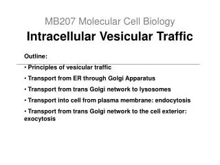
Microtubules 1) Organise the endoplasmic reticulum and the Golgi apparatus • Act as a “railroad” along which vesicles move from the periphery to he centre of the cell and back again • 3) Form the spindle apparatus in mitotic cells 4) Act as motile elements in cilia and flagella
Turnover of Microtubules • Microtubules are extremely dynamic structures. They have two distinct ends, called + and – and subunits are preferentially added to the plus end and lost from the – end. • In the non-motile interphase cell the – end of many microtubules is embedded in the microtubule organising centre which is located close to the Golgi apparatus. • Microtubules may be stabilised by binding of capping proteins to their + end.
To Summarise • Microtubules radiate from the microtubule organising centre • Vesicles may move along the microtubules in both directions – they act like an intracellular railroad • The rough endoplasmic reticulum is spread out by the microtubules like an umbrella on its spokes • Microtubules can interact with other cytoskeletal elements
Motile Cells While most cells in the body are fixed in place by attachments to each other and basement membranes, some, like neutrophils and macrophages remain motile. Free living single cells are generally motile and cell movement plays an important in early embryogenesis. Microfilaments and microtubules interact to control cell movement. This will be considered in more detail later on.
Intermediate Filaments 1) Form cables which stretch across the cell from desmosomes on one side to desmosomes on the other so giving strength to the cells 2) Hold the nucleus in place 3) May play a role in organising “permanent” cell extensions such as nerve axons 4) The nuclear lamina is formed by proteins called lamins whichare closely related to intermediate filament proteins
Continued Intermediate filamentsts are concentrated in the region round the nucleus and are responsible for holding the nucleus in place. Other intermediate filaments radiate to the cell surface and attach to junction complexes.
Organising tissues Cells are held together by cell adhesion molecules which may be single molecules or may be aggregates which form the different types of junction complex. The intracellular portion of junction complexes may interact with cytoskeletal elements or with cytosolic signalling proteins
Basal Lamina etc • In addition to the links between cells there are also links between cells and the extracellular matrix. In the centre of this photo is the basal lamina, above this are the epithelial cells, below it is the loose connective tissue of the sub-epithelium
References and further reading • This is a revision lecture so there is no specific reading list. However you should look back at your first year notes and at your favourite text book. Most figures here come from Alberts et al. but a few come from Lodish et al. These figures may be found on www.ncbi.nih.gov/books