The Eye
The Eye. Structures of the Eye-use handout. 1. Sclera -thick outer white portion of eye, maintains shape, protects. 1a. Cornea - clear, transparent, avascular layer, contacts are placed on top of this layer. 2. Choroid coat -contains blood vessels that nourish the eye.

The Eye
E N D
Presentation Transcript
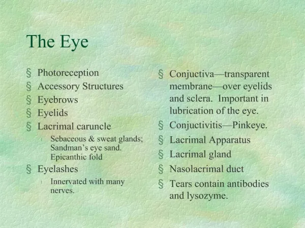
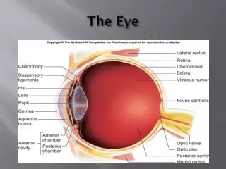
Structures of the Eye-use handout 1. Sclera-thick outer white portion of eye, maintains shape, protects. 1a.Cornea-clear, transparent, avascular layer, contacts are placed on top of this layer. 2. Choroid coat-contains blood vessels that nourish the eye. 2a. Ciliary body-helps control the lens. 2c.Iris-adjusts to allow light into the eye 3. Retina-at the back of the eye, contains photoreceptors that detect light(rods) and color (cones).
3a. Fovea centralis-concentration of rods and cones, most accutevision here. 3b. Optic disc-location where blood vessels and nerve enter and exit the eye, the blind spot! 3c. Optic nerve-carries message to the occipital lobe of brain. 5. Eyelid-provides protection for the eye. 7. Lens-adjusts to focus light onto the retina in the back of the eye 7a. Suspensory Ligaments-control the lens, attached to ciliary body. http://www.youtube.com/watch?v=RE1MvRmWg7I&feature=related
9. Pupil-opening in the central part of the eye, adjusted by the iris. 10. Anterior cavity-contains aqueous humor that nourishes and maintains shape of outer eye. 12.Vitreous humor-maintains shape of the eye, keeps pressure in the posterior cavity of the eye.
Accommodation http://www.youtube.com/watch?v=f2lwYBiy_wI
B. Disorders of the Eye 1.Myopia-nearsightedness, image is focused in front of the retina, eyeball is too long. Corrected with a concave lens. 2. Hypermetropia-farsightedness, image is focused behind the retina, eyeball is too short. Corrected by a convex lens. 3. Presbyopia-normal degeneration of focusing power as we age. Near point of vision increases and bifocals are needed.
4.Astigmatism-shape of cornea or lens is irregular so image is blurred. 5.Retinal detachment-retina separates from tunics of eye, vitreous humor decrease causes this or a blow to the eye and head, can cause blindness. 6. Color blindness- Inability to detect certain wavelengths of light, either red or green. a. red-green colorblindness -one cannot distinguish between red and green, one color will be detected. (red or green) b. sex linked trait carried by genes on x chromosomes.
7.Glaucoma- eye disease where there is an increase in pressure caused by aqueous humor. Leads to blindness if not treated. 8. Cataracts-clouding of lens by the buildup of protein