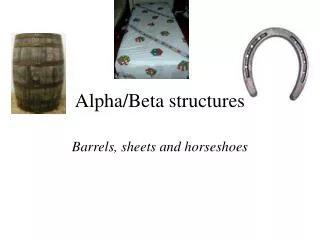
Beta structures
Beta structures. An awful lot of barrels. Functionally the most diversily populated group (antibodies, enzymes, transport proteins etc…) Second biggest group of protein domain structures (after a/b ). Common properties. Built up from four to over ten beta strands

Beta structures
E N D
Presentation Transcript
Beta structures An awful lot of barrels...
Functionally the most diversily populated group (antibodies,enzymes, transport proteins etc…) • Second biggest group of protein domain structures (after a/b)
Common properties • Built up from four to over ten beta strands • b strands are arranged in predominantly antiparallel fashion • Usually two beta sheets are formed, which pack each against other, resembling barrel or distorted barrel (=double b sandwich)
Up-and-down barrels • Simplest topology • Similar arrangement to TIM barrels, but without helices and all strands are antiparallel
Retinol-binding protein (rbp) • Retinol binds in the inside of barrel (typical for up-and-down barrels)
Retinol binding site in rbp • Hydrophobic part fits in a hydrophobic pocket • Hydroxyl group exposed to solvent OH
Alterating patterns in amino acid sequence of rbp • Hydrophobic amino acids are facing the core • Polar, charged and a few small hydrophobic are exposed to the solvent
Up-and-down barrels can contain more than 8 strands • Porin monomer from Rhodobacter has 14 b strands
b propeller in neuraminidase • Influenza virus protein, involved in virion release from cells • Tetrameric protein, one monomer consists of 6 up-and down b sheets • Builds a propeller-like structure
Active site in b-propeller proteins • On the top of propeller there are extensive loops • The loops form active site
g-crystallin • Found in lenses of your eyes • Each domain built from 2 greek key motifs • One connection across the barrel between two motifs
Evidence for two gene duplication events in g-crystallin evolution • Two domains have about 40% sequence identity • Two motifs within the domain share 20-30% sequence identity 1. 2. x 2 x 2
AH – CHOO ! Jelly-roll barrel in viruses • Very common in subunits of spherical viruses • Barrel is distorted and with helices instead of some loops • Example: Rhinovirus (common cold, that is)
Comparison of all those b-barrels Up-and-down g-crystallin-like jelly-roll
Yet another barrel – chymotrypsin fold • Present in chymotrypsin and all other serine proteases • Several non-protease proteins also contain similar fold • Six strands form the barrel
Structure of chymotrypsin Domain 1 Domain 2
Beta helix • Two different kinds – two-sheet helix and three-sheet helix • Both represent deviations from idealized structure with a single spiral-like strand
Sequence pattern in two sheet beta helix X9 X7 U8 U8 X7 • Gly-Gly-X-Gly-X-Asp-X-U-X • X=any amino acid • U=big hydrophobic, often Leu • Ca ions sit in between loops • Motif present in several bacterial proteases X9
Three sheet beta helix Unlike two-sheet beta helices, there are no repetitive sequence patterns
Structure of spider silk • All-beta fibrous protein • N- and C-terminal parts are variable • A large, up to 800 residues long central region is made from repeats: -(Ala)8-10-Gly-Gly-X-
Structure of spider silk Made up from beta sheets About 30% of beta sheets form microcrystals The rest of beta shets form a flexible matrix Soluble form of spider silk is a-helical! Beta sheets form upon spinning
Properties of spider silk • 5 times stronger than steel • Very elastic – can be stretched 3-4 times its original size without breaking • Lighter than cotton