Download
1 / 31
340 likes | 492 Views
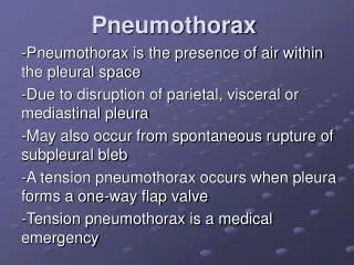
This chapter provides an in-depth exploration of pneumothorax, detailing the anatomic alterations of the lungs, including lung collapse and atelectasis. It categorizes pneumothorax into closed, open, and tension types, as well as traumatic, spontaneous, and iatrogenic origins. Key clinical manifestations, respiratory care protocols, and management strategies are discussed, emphasizing the importance of timely intervention to prevent severe complications. Radiologic findings further assist in the diagnosis and evaluation of this critical condition.

E N D
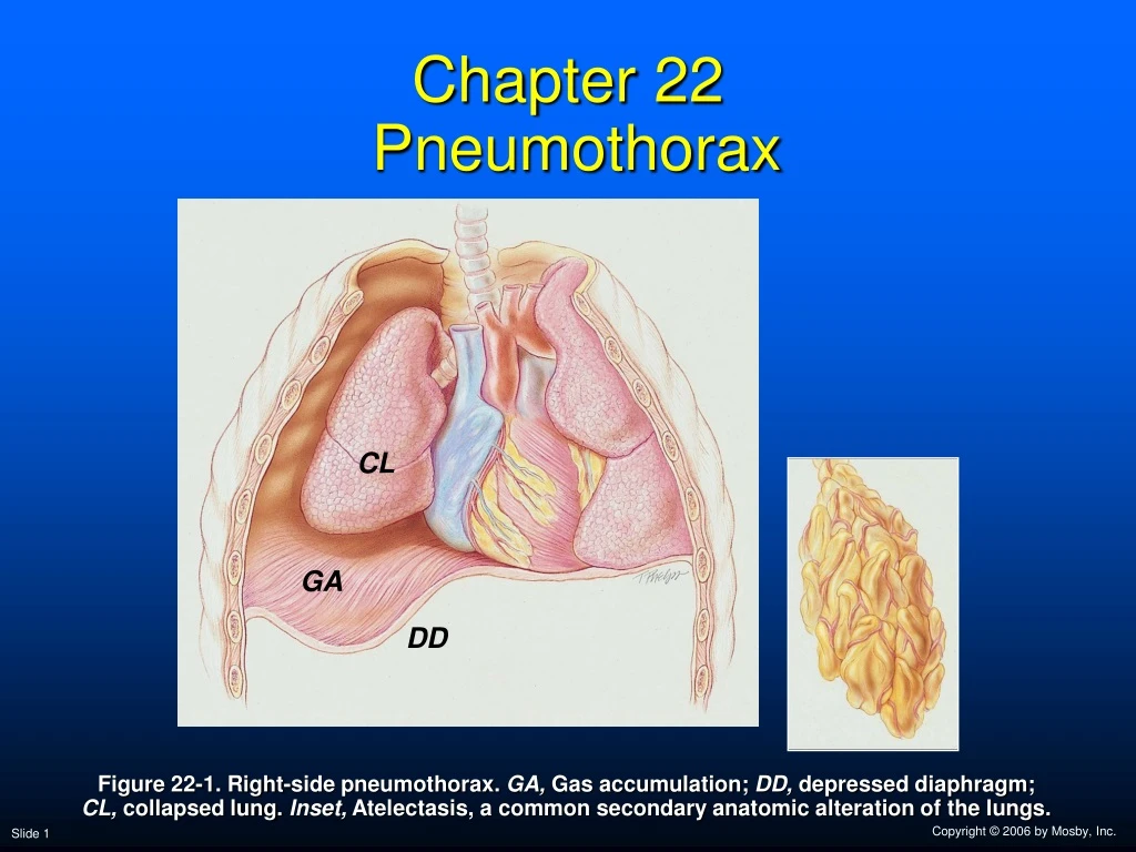
Chapter 22 Pneumothorax CL GA DD Figure 22-1. Right-side pneumothorax. GA, Gas accumulation; DD, depressed diaphragm;CL, collapsed lung. Inset, Atelectasis, a common secondary anatomic alteration of the lungs.
Anatomic Alterations of the Lungs • Lung collapse • Atelectasis • Chest wall expansion • Compression of the great veins and decreased cardiac venous return
Etiology—3 Ways • From the lungs through a perforation of the visceral pleura • From the surrounding atmosphere through a perforation of the chest wall and parietal pleura or, rarely, through an esophageal fistula or a perforated abdominal viscus • From gas-forming microorganisms in an empyema in the pleural space (rare)
Pneumothorax ClassificationsGeneral Terms • Closed pneumothorax • Open pneumothorax • Tension pneumothorax
Pneumothorax ClassificationsBased on Origin • Traumatic pneumothorax • Spontaneous pneumothorax • Iatrogenic pneumothorax
Figure 22-3. Closed (tension) pneumothorax produced by a chest wall wound.
Figure 22-4. Pneumothorax produced by a rupture in the visceral pleura that functions as a check valve.
Overview of the Cardiopulmonary Clinical Manifestations Associated with PNEUMOTHORAX The following clinical manifestations result from the pathophysiologic mechanisms caused (or activated) by Atelectasis (see Figure 9-7)—the major anatomic alterations of the lungs associated with pneumothorax (see Figure 22-1).
Clinical Data Obtained at the Patient’s Bedside Vital signs • Increased respiratory rate • Stimulation of peripheral chemoreceptors • Other possible mechanisms • Decreased lung compliance • Activation of the deflation receptors • Activation of the irritant receptors • Stimulation of the J receptors • Pain/anxiety • Increased heart rate, cardiac output, blood pressure
Clinical Data Obtained at the Patient’s Bedside • Cyanosis • Chest assessment findings • Hyperresonant percussion note over the pneumothorax • Diminished breath sounds over the pneumothorax • Tracheal shift • Displaced heart sounds • Increased thoracic volume on the affected side • Particularly in tension pneumothorax
Figure 22-6. Because the ratio of extrapulmonary gas to solid tissue increases in a pneumothorax, hyperresonant percussion notes are produced over the affected area.
Figure 22-7. Breath sounds diminish as gas accumulates in the intrapleural space.
Figure 22-8. As gas accumulates in the intrapleural space, the chest diameter increases on the affected side in a tension pneumothorax.
Clinical Data Obtained from Laboratory Tests and Special Procedures
Pulmonary Function Study: Lung Volume and Capacity Findings VT RV FRC TLC N or VC IC ERV RV/TLC% N
Arterial Blood Gases Small Pneumothorax • Acute alveolar hyperventilation with hypoxemia pH PaCO2 HCO3- PaO2 (Slightly)
Time and Progression of Disease Disease Onset Alveolar Hyperventilation 100 90 Point at which PaO2 declines enough to stimulate peripheral oxygen receptors 80 70 60 PaO2 PaO2 or PaCO2 50 40 30 PaCO2 20 10 0 Figure 4-2. PaO2 and PaCO2 trends during acute alveolar hyperventilation.
Arterial Blood Gases Large Pneumothorax • Acute ventilatory failure with hypoxemia pH PaCO2 HCO3- PaO2 (Slightly)
Time and Progression of Disease Disease Onset Alveolar Hyperventilation Acute Ventilatory Failure 100 Point at which disease becomes severe and patient begins to become fatigued 90 Point at which PaO2 declines enough to stimulate peripheral oxygen receptors 80 70 PaCO2 Pa02 or PaC02 60 50 40 30 PaO2 20 10 0 Figure 4-7. PaO2 and PaCO2 trends during acute ventilatory failure.
Oxygenation Indices QS/QT DO2 VO2 C(a-v)O2 Normal (severe) O2ER SvO2
Hemodynamic Indices (Large Pneumothorax) CVP RAP PAPCWP CO SV SVICI RVSWI LVSWI PVRSVR
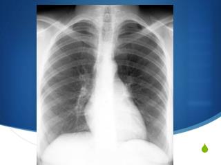
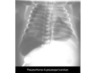
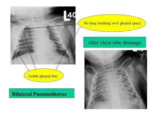
Radiologic Findings Chest radiograph • Increased translucency • Mediastinal shift to unaffected side in tension pneumothorax • Depressed diaphragm • Lung collapse • Atelectasis
Figure 22-9. Left-sided pneumothorax (arrows). Note the shift of the heart and mediastinum to the right away from the tension pneumothorax.
B A Figure 22-10. A, Development of a small tension pneumothorax in the lower part of the right lung (arrow). B, The same pneumothorax 30 minutes later. Note the shift of the heart and mediastinum to the left away from the tension pneumothorax. Also note the depression of the right hemidiaphragm (arrow).
General Management of Pneumothorax • >20%—gas should be evacuated • Negative pressure—5 to 12 cm H2O • Should not exceed negative 12 cm H2O
General Management of Pneumothorax Respiratory care treatment protocols • Oxygen therapy protocol • Hyperinflation therapy protocol • Mechanical ventilation protocol
General Management of Pneumothorax PLEURODESIS • Chemical or medication injected into the chest cavity • Talc • Tetracycline • Bleomycin sulfate • Produces inflammatory reaction between lungs and inner chest cavity • Causes lung to stick to chest cavity