Download
1 / 10
100 likes | 117 Views
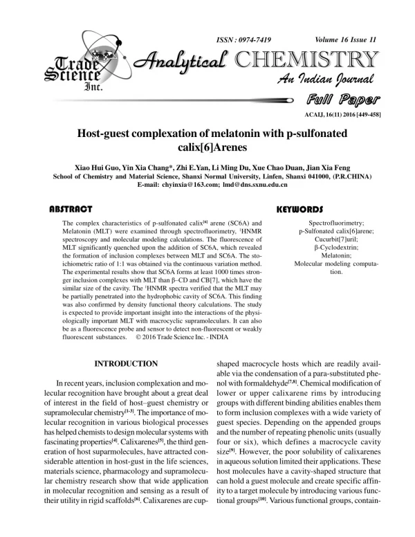
The complex characteristics of p-sulfonated calix [6] arene (SC6A) and Melatonin (MLT) were examined through spectrofluorimetry, 1HNMR spectroscopy and molecular modeling calculations. The fluorescence of MLT significantly quenched upon the addition of SC6A, which revealed the formation of inclusion complexes between MLT and SC6A. The stoichiometric ratio of 1:1 was obtained via the continuous variation method. The experimental results show that SC6A forms at least 1000 times stron- ger inclusion complexes with MLT than u201au00f1CD and CB[7], which have the similar size of the cavity. The 1HNMR spectra verified that the MLT may be partially penetrated into the hydrophobic cavity of SC6A. This finding was also confirmed by density functional theory calculations.

E N D
id5087703 pdfMachine by Broadgun Software - a great PDF writer! - a great PDF creator! - http://www.pdfmachine.com http://www.broadgun.com Volume 16 Issue 11 ISSN : 0974-7419 Analytical Analytical CHEMISTRY Analytical Analytical CHEMISTRY An Indian Journal Full Paper Full Paper ACAIJ, 16(11) 2016 [449-458] Host-guest complexation of melatonin with p-sulfonated calix[6]Arenes Xiao Hui Guo, Yin Xia Chang*, Zhi E.Yan, Li Ming Du, Xue Chao Duan, Jian Xia Feng School of Chemistry and Material Science, Shanxi Normal University, Linfen, Shanxi 041000, (P.R.CHINA) E-mail: chyinxia@163.com; lmd@dns.sxnu.edu.cn ABSTRACT KEYWORDS The complex characteristics of p-sulfonated calix[6]arene (SC6A) and Melatonin (MLT) were examined through spectrofluorimetry, 1HNMR spectroscopy and molecular modeling calculations. The fluorescence of MLT significantly quenched upon the addition of SC6A, which revealed the formation of inclusion complexes between MLT and SC6A. The sto- ichiometric ratio of 1:1 was obtained via the continuous variation method. The experimental results show that SC6A forms at least 1000 times stron- ger inclusion complexes with MLT than â–CD and CB[7], which have the similar size of the cavity. The 1HNMR spectra verified that the MLT may be partially penetrated into the hydrophobic cavity of SC6A. This finding was also confirmed by density functional theory calculations. The study is expected to provide important insight into the interactions of the physi- ologically important MLT with macrocyclic supramoleculars. It can also be as a fluorescence probe and sensor to detect non-fluorescent or weakly fluorescent substances. 2016 Trade Science Inc. - INDIA Spectrofluorimetry; p-Sulfonated calix[6]arene; Cucurbit[7]uril; â-Cyclodextrin; Melatonin; Molecular modeling computa- tion. INTRODUCTION shaped macrocycle hosts which are readily avail- able via the condensation of a para-substituted phe- nol with formaldehyde[7,8]. Chemical modification of lower or upper calixarene rims by introducing groups with different binding abilities enables them to form inclusion complexes with a wide variety of guest species. Depending on the appended groups and the number of repeating phenolic units (usually four or six), which defines a macrocycle cavity size[9]. However, the poor solubility of calixarenes in aqueous solution limited their applications. These host molecules have a cavity-shaped structure that can hold a guest molecule and create specific affin- ity to a target molecule by introducing various func- tional groups[10]. Various functional groups, contain- In recent years, inclusion complexation and mo- lecular recognition have brought about a great deal of interest in the field of host–guest chemistry or supramolecular chemistry[1-3]. The importance of mo- lecular recognition in various biological processes has helped chemists to design molecular systems with fascinating properties[4]. Calixarenes[5], the third gen- eration of host suparmolecules, have attracted con- siderable attention in host-gust in the life sciences, materials science, pharmacology and supramolecu- lar chemistry research show that wide application in molecular recognition and sensing as a result of their utility in rigid scaffolds[6]. Calixarenes are cup-
Host-guest complexation of melatonin with p-sulfonated calix[6]Arenes . 450 ACAIJ, 16(11) 2016 Full Paper Full Paper MLT SC6A CB[7] ²-CD Scheme 1 : The structure of molecules are melatonin, p-sulfonated calix[6]arene, cucurbit[7]uril and â–cyclodextrin successively ing dialkylamino groups, phosphonic acid groups, carboxyl groups, sulfonic groups and so on, could be introduced to either the upper or the lower rim of the ‘cup’, which could change the affinity of these cyclooligomers towards target molecules or increase the solubility of the calixarenes[11-13]. Studies have investigated p-Sulfonated calix[6]arene (SC6A) (scheme 1), which have flex- ible and often poorly defined cavities that bind posi- tively charged species. SC6A are regarded as prom- ising water-soluble hosts[14]for quaternary ammo- nium ions[15,16], trimethylammonium cations[17-19], dyes[20,21], native amino acids[22,23] and small neutral organic molecules[24]. Furthermore, SC6A have been applied in the improvement of solubility and stabil- ity of drugs and enzyme mimics[25-29]. Melatonin (MLT) (scheme 1) is well known as a paracrine hormone that is secreted in a cyclic man- ner by the pineal gland[30]. In mammals, the pineal gland is believed to be the major source of circulat- ing melatonin[31]. It detoxifies a variety of free radi- cals (OH), peroxynitrite anion, singlet oxygen, and nitric oxide[32]. Melatonin controls biological rhythms, pigment metabolism, immune response, me- tabolism of free radicals, monitoring of mood and sleep, cell proliferation and differentiation[33]. MLT can perform as a valuable drug for prevention or cure of several diseases; its present breadth of ap- plications, ranging from sleep-induction and antiageing action to cancer therapy, could be further extended by several clinical or pre-clinical studies in progress[34]. Over the past years, interactions between MLT and supramolecules or host–guest molecules have been widely studied. Ernane et.al[35], discovered a spectrofluorimetric method for the determination of MLT through the interaction with lipid bilayers. Hany et.al[36], investigated some new spectrofluorimetric methods for determination of MLT in the presence of N-{2-[1-({3-[2-(acetylamino)ethyl]-5-methoxy- 1H-indol-2-yl}methyl)-5-methoxy-1H-indol-3-yl]- ethyl}acetamide: a contaminant in commercial me- latonin preparations, MLT was determined in labo- ratory prepared mixtures containing different per- centages of compound. However, to the best of our knowledge, the supramolecular interactions between Analytical Analytical CHEMISTRY Analytical Analytical CHEMISTRY An Indian Journal
ACAIJ, 16(11) 2016 Yin Xia Chang et al. 451 Full Paper Full Paper SC6A and MLT were determined by spectrofluo- rometry have not been reported. In our experiment, MLT have highly fluorescent nature and efficient fluorescence quenching prop- erty upon binding with SC6A by fluorescence titra- tions, A gradual decrease in the fluorescence inten- sity was observed with the SC6A concentration in- creased. Temperature-dependent inclusion constants were also obtained, 1HNMR spectra and molecular modeling analyses were performed to investigate the possible mechanism of the binding reaction. Moreover, a comparative study of the complex- ation behavior of fluorescent MLT with cucurbit[7]uril (CB[7]) and â-Cyclodextrin (â–CD) (scheme 1) has been carried out on the basis of re- sults obtained by spectrofluorimetry, 1HNMR spec- troscopy and molecular modeling calculations. (China). Its purity was 99.5%. The stock solution of 1.0×10⁻³ mol L-1 was prepared by directly dissolv- ing in double-distilled water. SC6A was purchased from TCI Chemical Industry Co, Ltd (Shanghai, China). SC6A stock solution of 1.0×10⁻³ mol L-1 were prepared respectively in a 100 mL volumetric flask. -Cyclodextrin (Sinopharm Chemical Reagent Shanghai Co., Ltd.) was recrystallized twice from water and dried under a vacuum at 95 ºC for 24 h prior to use. CB[7] was prepared and characterized according to recently reported procedures[37]. Work- ing solutions were obtained by dilution of the stock solution. The standard MLT solution was found stable over time during the experiment, its fluores- cence intensity was not change. The stock solutions were kept at 4°C degree. The working solution was prepared freshly. The SC6A stock standard solutions were stable for several weeks at room temperature. A Britton-Robinson (BR) buffer solution (pH 2.00– 12.00) was prepared using 0.04 mol L”1 boric acid, acetic acid and phosphoric acid, and then was ad- justed to accurate values by using 0.2 mol L”1 so- dium hydroxide. Experimental procedure A total of 1.0 mL of 1.0×10⁻⁴ mol L-1 MLT solu- tion was transferred into a 10 mL volumetric flask, and an appropriate amount of 1.0×10⁻⁴ mol L-1 SC6A was added. The pH was controlled by 1.0mL of Britton-Robinson buffer solutions. The mixed solu- tion was diluted to the final volume with distilled water and shaken thoroughly, then equilibrated for 15 min at room temperature. The fluorescence in- tensity values of the experimental and blank solu- tions (FMLT) were measured using at 358 nm an ex- citation wavelength of 227 nm. EXPERIMENTAL Apparatus Fluorescence spectra and measurements were obtained using a Agilent Technologies Cary Eclipse Fluorescence spectrofluorometer equipped with 150 W xenon lamp (Japan). The slit widths of both the excitation and emission monochromators was set at 5 nm. The fluorescence spectra were recorded at a scan rate of 600 nm min”1. All measurements were performed in a standard 10 mm path-length quartz cell set to a temperature of 25.0±0.5°C. The pH val- ues were measured using a pHS-3TC digital preci- sion pH meter (Shanghai, China). In the experiment, the temperatures were controlled by using a thermostated cell holder and a thermostatically con- trolled water bath. 1HNMR spectra was recorded using a Bruker DRX-600MHz spectrometer (Swit- zerland). Molecular modeling calculations were optimized at the B3LYP/6-31G(d) level of density functional theory with the Gaussian 03 program. Reagents All reagents used were of analytical reagent grade or the best grade available commercially, and double-distilled water was used throughout the pro- cedures. The MLT used in the experiment were ob- tained from the National Institute of Metrology RESULTS AND DISCUSSION Fluorescence quenching of the SC6A-MLT com- plex Aqueous solutions of MLT have strong fluores- cence in aqueous solution and the maximum excita- tion and emission wavelengths were located at 227 and 358 nm, respectively. However, addition of SC6A brought a markedly decrease in MLT fluores- Analytical Analytical CHEMISTRY Analytical Analytical CHEMISTRY An Indian Journal
Host-guest complexation of melatonin with p-sulfonated calix[6]Arenes . 452 ACAIJ, 16(11) 2016 Full Paper Full Paper Figure 1 : The fluorescence spectra of MLT in different concentrations of SC6A in britton–robinson buffer solu- tions. The concentrations of SC6A(10-4mol L-1) (1) 0; (2) 0.3; (3) 0.5; (4) 0.7; (5) 0.9; (6) 1.0; (7) 1.2; (8) 1.5; (9) 1.7; (10) 2.0; CMLT=1.0×10⁻⁴mol L-1 cence intensity. These modifications of the features of the fluorescence spectra were considered to be a result of inclusion complex formation between MLT and SC6A. An interaction between MLT and SC6A was con- firmed by flurescence spectra when the initial concetration of MLT was 1.0×10⁻⁴ mol L-1. Figure 1 shows the fluorescence spectra of MLT in the ab- sence and presence of SC6A. Fluorescence inten- sity gradually decreased with increased SC6A con- centration. The temperature and pH of the experiment were optimized. In Britton–Robinson buffer solutions with pH 7.4 and at room temperature, the fluorescence of MLT was significantly quenched upon a certain amount of SC6A. Therefore, a pH of 7.4 was used for all subsequent experiments and room tempera- ture was selected as the standard reaction condi- tion. Stoichiometry and association constant of the in- clusion complex Inclusion capacity of a host in relation to spe- cific guest was described as the inclusion formation constant (K). Under the optimum experimental con- ditions, with 1:1 MLT-SC6A complex, K could be obtained by the following calculation: SC6A+MLTSC6A-MLT SC6A-MLT C K = C Where CMLT, CSC6A, and CMLT–SC6A are presented as equilibrium concentrations. Thus, K value can be determined by the typical double reciprocal or Benesi-Hildebrand plots[38]. 1 1 ( ) F F F F KC Where F is the observed fluorescence intensity when each corresponding SC6A concentration was tested, F0 is the MLT fluorescence intensity without SC6A addition, and F” is the enhancement once all MLT complexs are formed. A good linear relationship was obtained when 1/(F-F0) was plotted against 1/CSC6A, which supports the existence of a 1:1 complex(Figure 2). The bind- ing constants of the MLT and SC6A complexes at pH 7.4 were determined to be 4.97×10⁴ M-1 in the presence of SC6A. The value was determined by dividing the intercept by the slope of the Y corre- sponding lines. Generally, the binding stoichiometry of MLT with SC6A was determined by Job’s plots and was found to be 1:1(Figure 3). The maximum of the relative fluorescence intensity was at a mol fraction of 0.5, which confirmed the formation of a 1:1 ratio com- (2) C SC6A MLT 1 (3) F F SC6A 0 0 0 (1) Analytical Analytical CHEMISTRY Analytical Analytical CHEMISTRY An Indian Journal
ACAIJ, 16(11) 2016 Yin Xia Chang et al. 453 Full Paper Full Paper Figure 2 : The relationship of (F-F0)-1 with SC6A-1, T=298K, C(MLT)=10-4M, pH=7.4 Figure 3 : Job’s plot for the complex of MLT with SC6A in Britton-Robinson buffer solution (pH 7.4) at 25!.([MLT]+[SC6A])=1.0×10⁻⁵ mol L-1 Figure 4 : The fluorescence spectra of MLT in different concentrations of â–CD in Britton–Robinson buffer solutions. The concentrations of â–CD (10-4 mol L-1) (1) 0; (2) 0.5; (3) 1.0; (4) 1.5; (5)2.0; (6) 3.0; (7) 4.5; CMLT=1.0×10⁻⁴mol L-1 Analytical Analytical CHEMISTRY Analytical Analytical CHEMISTRY An Indian Journal
Host-guest complexation of melatonin with p-sulfonated calix[6]Arenes . 454 ACAIJ, 16(11) 2016 Full Paper Full Paper Figure 5 : 1HNMR spectra (600 MHz) of (a) MLT (b) SC6A-MLT complex (c) â–CD-MLT complex and (d) CB[7]- MLT complex in D2O plex[39]. It is well known that the complexation between the guest and the calixarene are formed by weak forces including hydrogen bonding, p–p interaction, electrostatic interaction, hydrophobic interaction, dipole–dipole or van der Waals[44]. Generally, in the process of the formation of the inclusion complex, the key force depends on the structure, the charge, functional group of guest and host. SC6A has an up- per laced with six hydroxyl groups and a socially wider rim with modified sulfonic groups, and in- dole benzene ring of MLT is located in the vicinity of a carbonyl-laced portal. The formation of hydro- gen bond between hydrogen at N atom of indole ring and the sulfonic groups may cause the changes of the geometric configuration of indole ring. The carbanyl group and hydroxy of SC6A may form hydrogen bond. The NH of aliphatic chain is likely to form hydrogen bond with the hydroxy of SC6A. Hydro- gen bonding interaction and the electrostatic inter- action lead to the formation of host-guest inclusion complex. This state results in a less polar microen- vironment which, in turn, leads to fluorescence quenching. These results are consistent with the fore- going discussion. Comparison of the Photophysical Properties upon Complexation with SC6A, CB7 and â–CD. David et.al[45]. obtained the 1:1 complex forma- tion between MLT and â–CD from the 1HNMR spec- 1HNMR spectroscopy and molecular modeling calculation The formation of SC6A-MLT inclusion com- plexes in aqueous solution was confirmed using 1HNMR Spectroscopy(Figure 5b). Compared with the proton resonances of the unbound MLT molecules (Figure 5a), the signals from H1, H2, H3, H4, H5, H6, H7, and H9 protons of the bound MLT signifi- cantly shifted upfield. Especially, the singlets for H3, H4 protons inside the SC6A cavity should be sensi- tive to the changed environment after the complex- ation were observed clearly. This behavior is characteristic of this part of the MLT molecule en- capsulated in the SC6A cavity. Furthermore, sev- eral protons chemical shifts of MLT were changed. It confirmed that the complex was formed. These results are consistent with the previous fluorescence quenching. Molecular modeling calculations were optimized at the B3LYP/6-31G[40] lever of density functional theory[41,42] using the Gaussian 03 program[43]. Mo- lecular mechanics was simulated to obtain the opti- mized conformation of the host-guest complex (Fig- ure 6). The SC6A-MLT molecular models are shown in Figure 6a. In the energy minimized structure, the complexation reactions MLT with SC6A are very sensitive since the high flexibility of SC6A. Analytical Analytical CHEMISTRY Analytical Analytical CHEMISTRY An Indian Journal
ACAIJ, 16(11) 2016 Yin Xia Chang et al. 455 Full Paper Full Paper troscopy, calorimetric and solubility measurements, and mass spectrometry. The inclusion constants for these 1:1 complexes were determined to be 48.0 M- 1 in the presence of â–CD. The point to note here is that the binding constant significantly less than the binding constants of the SC6A inclusion complexes. In our study, the supramolecular interaction between MLT and â–CD has been studied using fluorometric method. Figure 4 showed the fluorescence spectra of the MLT in the absence and presence of â–CD. The fluorescence intensity of the MLT significantly decreased upon the addition of â–CD. The fluores- cence quenching of SC6A was more significant than that of â–CD at the same MLT concentration. Mean- while, we examined the interaction of CB[7] with MLT by fluorometric method and the system has no the effect of fluorescence change. The mechanisms of the inclusion process of MLT with â–CD and CB[7] have also been discussed by the 1HNMR and molecular modeling calculation. A rough estimation of the hydrodynamic molecu- lar geometric dimensions can be obtained by con- sidering the complex as an effective sphere and the hydrodynamic diameter similar to the inside diam- eters of the SC6A (0.76 Å), CB[7] (0.73 Å) and â– CD (0.78Å). The formation of SC6A-MLT, CB[7]- MLT, and â–CD-MLT inclusion complexes in aque- ous solution was confirmed using 1HNMR spectros- copy experiments were carried out in D2O at room temperature. Figure 6 also shows the 1HNMR spec- tra of a 1:1 host-guest complex betweeen MLT and the two macrocyclic host molecules. Compared with the proton resonances of the free MLT molecules(Figure 5a), the chemical shift of MLT protons changed after complexation with mac- rocyclic host molecules (Figure 5). The signals from H3 and H4 protons of the bound â–CD significantly shifted upfield, this behavior is characteristic of this part of the indole benzene ring of the MLT molecule encapsulated in the â–CD cavity. The chemical shift of the H1, H2, H5, H6, H7 and H9 protons of the â– CD-MLT is practically unchanged, which indicates the protons of this part of the molecule located just outside the cavity of the â–CD host. The resonance of protons H3 of CB[7]-MLT experienced a slightly up-field shift, indicating this part of the molecule was located just inside the carbonyl portal of the CB[7] host. The chemical shift of the H2, H3, H4, H5, H6, H7 and H9 protons of the CB[7]-MLT is practically unchanged, indicating this part of the molecule was located just outside the carbonyl por- tal of the CB[7] host. The MLT molecules immersed in the cavity of SC6A, which is expected to enter CB[7] and â–CD cavity. The result confirmed the partial inclusion of MLT in the hydrophobic cavity of â–CD, and CB[7] using the Gaussian 03 program, Respectively(Figure 6). The restriction of the conformational mobility is not compensated by the release of water molecules from the MLT molecules and CB[7] a slight rise in the chemical shift is observed for the weaker-bound MLT-CB[7] complexes, hydrogen bonds[46] and elec- trostatic interactionsbetween both molecules are weak. The polar carbonyl groups at each portal of CB[7] and their simultaneous interactions with the protonated amino groups are responsible for this ef- fect. The NH of aliphatic chain is located in the vi- cinity of a carbonyl-laced portal, the methoxy group is located in the vicinity of the other carbonyl-laced portal. The partial immersion of MLT in the hydro- phobic cavity via portals of CB[7] attributed to hy- drogen bonding. The hydroxyl groups located at both rims of â-CD are likely to form hydrogen bonds with the amino groups and the NH of the indole ring. Hy- drogen bonding interaction may lead to the forma- tion of host-guest inclusion complex. Because of the excellent matched size and morphology, benzene ring residues were enclosed in the cavity of â–CD more tightly than CB[7] in aqueous solution. These re- sults are consistent with the trend observed in 1HNMR spectra and the previous fluorescence phe- nomenon. Generally speaking, SC6A forms at least 1000 times stronger inclusion complexes with MLT than â–CD and CB[7]. For potential practical applica- tions, it is important to note that a virtually quantita- tive complexation of MLT can be readily achieved even with small amounts of SC6A. This is due to the high binding constants. From a photophysical point of view, the addition of both host molecules results in a fluorescence quenching of MLT. Because of the more flexible conformation of SC6A â–CD Analytical Analytical CHEMISTRY Analytical Analytical CHEMISTRY An Indian Journal
Host-guest complexation of melatonin with p-sulfonated calix[6]Arenes . 456 ACAIJ, 16(11) 2016 Full Paper Full Paper a(1) a(2) b(1) b(2) c(1) c(2) Figure 6 : Lowest energy structure of complex using ball and stick model determined by molecular dynamics simulation with direct minimization for the rendering of atoms. (a) MLT and SC6A complex, (b) MLT and â–CD complex, (c) MLT and CB[7] complex. (1) profile (2)above CB[7], the hydrophobic segments of the benzene ring of SC6A may be the most easily adsorbed to MLT molecule. (see Figure 6). The dramatic fluorescence quenching provides strong evidence that inclusion complexes are indeed formed between MLT and SC6A. Analytical Analytical CHEMISTRY Analytical Analytical CHEMISTRY An Indian Journal
ACAIJ, 16(11) 2016 Yin Xia Chang et al. 457 Full Paper Full Paper CONCLUSIONS [4] (a) E.A.Meyer, R.K.Castellano, F.Diederich; Angew.Chem.Int.Ed., 42, 1210 (2003); (b) J.Joseph, N.V.Eldho, D.Ramaiah; J.Phys.Chem, 107, 4444 (2003); (c) Z.Xu, R.J.Song, H.Moon, J.Y.Lee, J.Yoon; Org.Biomol.Chem., 9, 8340 (2011). Q.Li, D.Guo, H.Qian, Y.Liu; Eur.J.Org.Chem., 21, 3962-3971 (2012). Y.Wu, X.Ni, L.Mou, C.Jin, C.Redshaw, T.Yamato; Supramol.Chem., 27(7-8), 501-507 (2015). C.D.Gutsche; Acc.Chem.Res., 16, 161–170 (1983). V.Bôhmer; Angew.Chem.Int.Ed., 34, 713–745 (1995). G.Nives, B.Natasa, T.Renato, F.Leo, T.Vladislav; Supramol.Chem., 23, 389-397 (2011). [10] M.Chen, T.S.Shang, J.Chem.Thermodyn., 43, 88–93 (2011). [11] S.Shinkai, S.Mori, H.Koreishi, T.Tsubaki, O.Manabe; J.Am.Chem.Soc., 108, 2409–2416 (1986). [12] I.Matulková, J.Rohovec; Polyhedron., 24, 311–317 (2005). [13] T.Oshima, R.Saisho, K.Ohe, Y.Baba, K.Ohto; React.Funct.Polym., 69, 105–110 (2009). [14] Y.Y.Zhou, Q.Lu, C.Liu, S.K.She, L.Wang; Ana.J.Chim.Acta., 552, 152–159 (2005). [15] A.Arena, N.Donato, G.Saitta, A.Bonavita, G.Rizzo, G.Neri; Sensors Actuators B Chem., 145, 488–494 (2010). [16] R.Arnecke, V.Böhmer, R.Cacciapaglia, A.Cort, L.Mandolini; Tetrahedron., 53, 4901–4908 (1997). [17] B.Nimse, J.Kim, K.Song, J.Kim, J.Lee, V.Nguyen, V.Ta, T.Kim; Tetrahedron Lett., 52, 3751–3755 (2011). [18] G.Arena, S.Gentile, F.Gulino, D.Sciotto, C.Sgarlata; Tetrahedron Lett., 45, 7091–7094 (2004). [19] T.Komura, T.Yamaguchi, K.Kura, J.Tanabe; J.Electroanal Chem., 523, 126–135 (2002). [20] A.Yilmaz, E.Yilmaz, M.Yilmaz, R.Bartsch; J.Hazard Mater., 74, 54–59 (2007). [21] E.Akceylan, M.Bahadir, M.Yýlmaz; J.Hazard Mater., 162, 960–966 (2009). [22] E.P.Silva, A.C.Shahgaldian; Int.J.Pharm., 273, 57– 62 (2004). [23] M.Michele, H.Andreas, J.Am.Soc.Mass.Spectrom., 13, 964–974 (2002). [24] G.Arena, A.Contino, F.G.Gulino, A.Magri, D.Sciotto, R.Ungaro; Tetrahedron Lett., 41, 9327–9330 (2000). [25] W.Yang, M.M.Villiers; Eur.J.Phanrm.Biopharm., 58, 629–636 (2004). The results obtained from the spectrofluorimetric studies indicate that both SC6A form inclusion com- plexes with MLT, particularly strong for the interac- tion MLT-SC6A complex (K>104 M-1). According to the data, SC6A can react with MLT to from 1:1 inclusion complex. The association constants of the complexes formed between the host and the guest were calculated. The interaction mechanism was confirmed via the 1HNMR spectrum. The interac- tion models of the supramolecular complexes were established through theoretical calculations. The comparsion of the complexation behavior of MLT toward SC6A with that toward â–CD and CB[7] re- veals that although all hosts have a simliar hydro- phobic cavity, additional recognition elements such as sulfonic groups receptor sites at the portals of the cavity are quintessential to promote a strong and se- lective binding of MLT, which may affect the fluo- rescence intensity of MLT. The possible complexation mechanism for MLT and SC6A may involve hydrophobic and electro- static interaction. This study can be used as a fluo- rescence probe and sensor to detect non-fluorescent or weakly fluorescent substances. This work will be helpful to provide useful information for appro- priately understanding of the drug design and phar- maceutical research. Related studies are currently underway. [5] [6] [7] [8] [9] J.Liu, G.W.Diao; ACKNOWLEDGEMENTS This work was supported by the National Natu- ral Science Foundation of China (No. 81402895). Helpful suggestions by anonymous referees are also gratefully acknowledged REFERENCES B.Carlito; [1] C.A.Schalley; Int.J.Mass.Spectrom, 194, 11–19 (2000). [2] A.Kumar, S.S.Sun, A.J.Lees; Coordin.Chem.Rev., 252, 922–939 (2008). [3] Z.Garaiova, M.A.Mohsin, F.G.Banica Vargova´, T.Hianik; Bioelectrochemistry, 87, 220–225 (2012). Analytical Analytical CHEMISTRY Analytical Analytical CHEMISTRY An Indian Journal
Host-guest complexation of melatonin with p-sulfonated calix[6]Arenes . 458 ACAIJ, 16(11) 2016 Full Paper Full Paper [26] H.L.Liu, T.T.Pang, L.M.Du, Y.Zhao, X.Jing, F.H.Yao, Y.L.Fu; Spectroscopy Lett., 47, 306–313 (2014). [27] T.M.Yao, Z.F.Ye, L.Wang, J.Y.Gu, S.D.Yao, X.F.Shi; Spectrochim.Acta Part A., 58, 3033–3038 (2002). [28] T.T.Pang, H.L.Liu, L.M.Du, Y.X.Chang, Y.L.Fu; J.Fluoresc., 24, 143–152 (2014). [29] Y.Zhou, Q.Lu, C.Liu, S.She, L.Wang; Spectrochim.Acta A., 64, 748–756 (2006). [30] Bj.Teubner,; D.A.Freeman; J.Neuroendocrinol., 19(2), 102-108 (2007). [31] R.J.Reiter; Endocr.Rev., 12, 151 (1991). [32] D.X.Tian, L.C.Manchester, B.F.Plummer, J.Limson, S.T.Weintraub, W.Qi; Free Radical Bio.Med., 29(11), 1177-1185 (2000). [33] I.Kvetnoy, I.Ingel, T.Kvetnaia, N.Malinovskaya, S.Rapoport, N.Raikhlin, A.Trofimov, V.Yuzhakov; Neuroendocrinol Lett., 23, 121 (2002). [34] R.J.Reiter, P.J.Robinson, Bantam Books: New York., 12(10), 735-736 (1995). [35] E.J.X.Costa, C.S.Shida, M.H.Biaggi, A.S.Ito, M.T.Lamy-Freund; FEBS Lett., 416, 103-106 (1997). [36] H.W.Darwish, M.I.Attia; Darwish and Attia Chem- istry Central Journal., 6, 36 (2012). [37] J.Kim, I.S.Jung, S.Y.Kim; J.Am.Chem.Soc., 122, 540 (2000). [38] C.F.Li, L.M.Du, H.M.Zhang; Spectrochim.Acta Part A: Molecular and Biomolecular Spectroscopy., 75, 912-917 (2010). [39] H.W.Gibson, H.Wang, C.Slebodnick, J.Merola, W.Kassel, A.L.Rheingold; J.Org.Chem., 72, 3381– 3393 (2007). [40] C.Lee, W.Yang, R.G.Parr; Phys.Rev.B., 37, 785– 789 (1988). [41] A.D.Becke; J.Chem.Phys., 88, 2547–2553 (1988). [42] A.D.Becke; Phys.Rev.A., 38, 3098–3100 (1988). [43] M.J.Frisch, et al; Gaussian 03, Revision C.02, Gaussian, Inc., Wallingford, CT (2004). [44] Y.Inoue, Y.L.Liu, H.Tong, B.J.Shen, D.S.Jin; J.Am.Chem.Soc., 115, 10637–10644 (1993). [45] D.Bongiorno, L.Ceraulo, A.Mele, W.Panzeri, A.Selva,V.T.Liveri; Carbohyd Res., 337, 743–754 (2002). [46] V.Socorro, C.Mercedes, Supramol.Chem., 19, 95-106 (2007). R.J.Reiter, N.R.David; Analytical Analytical CHEMISTRY Analytical Analytical CHEMISTRY An Indian Journal
![THE STUDY OF SPECTROSGRAPHIC PROPERTIES AND MOLECULAR RECOGNITION OF CALIX[n]ARENES](https://cdn0.slideserve.com/327494/the-study-of-spectrosgraphic-properties-and-molecular-recognition-of-calix-n-arenes-dt.jpg)


![The Synthesis of Sulfo Derivatives of Calix[4]arenes](https://cdn1.slideserve.com/3415659/slide1-dt.jpg)