Download
1 / 40
440 likes | 744 Views
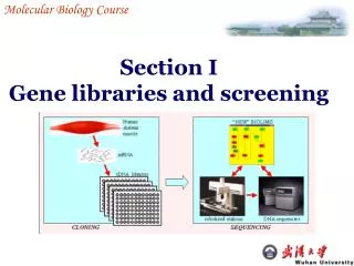
Molecular Biology Course. Section I Gene libraries and screening. Gene libraries and screening. I 1 Genomic libraries I 2 cDNA libraries I 3 Screening procedures. Gene libraries and screening. I 1 Genomic libraries. I 1-1 Representative gene libraries I 1-2 Size of library

E N D
Molecular Biology Course Section IGene libraries and screening
Gene libraries and screening I1 Genomic libraries I2 cDNA libraries I3 Screening procedures
Gene libraries and screening I1 Genomic libraries I1-1 Representative gene libraries I1-2 Size of library I1-3 Genomic DNA I1-4 Vectors
I1 Genomic libraries Gene library:a collection of different DNA sequence from an organism, each of which has been cloned into a vector for ease of purification, storage and analysis. Genomic libraries (made from genomic DNA) Gene library cDNA libraries (made from cDNA- copy of mRNA)
I1 Genomic libraries I1-1 Representative gene libraries --- Contain all the original sequences Missing original sequence • Certain sequences have not been cloned. • Example: repetitive sequences lacking restriction sites Too long for the vector used 2. Library does not contain sufficient clones
I1 Genomic libraries I1-2 Size of library(ensure enough clones) must contain a certain number of recombinants for there to be a high probability of it containing any particular sequence The formula to calculate the number of recombinants: ln (1-P) N = ln (1-f) P: desired probability f : the fraction of the genome in one insert
I1 Genomic libraries For example :for a probability of 0.99 with insert sizes of 20 kb these values for the E.coli (4.6×10⁶ bp) and human (3×10⁹ bp) genomes are : N E.coli= = 1.1 ×10³ ln( 1-0.99) ln[1-(2×10⁴/4.6×106)] ln(1-0.99) Nhuman= = 6.9 ×10⁵ ln[1-(2 ×10⁴/3 ×109)] These values explain why it is possible to make good genomic libraries from prokaryotes in plasmids where the insert size is 5-10kb ,as only a few thousand recombinants will be needed.
I1-3 Genomic DNA libraries Purify genomic DNA Fragment this DNA : physical shearing and restriction enzyme digestion I1 Genomic libraries eukaryotes prokaryotes Clone the fragments into vectors
I1 Genomic libraries To make a representative genomic libraries , genomic DNA must be purified and then broken randomly into fragments that are correct in size for cloning into the chosen vector. Purification of genomic DNA : Eukaryotes:preparecell nuclei removeprotein, lipids and other unwanted macro- molecules by protease digestion and phase extraction. Prokaryotes:extracted DNA directly from cells
I1 Genomic libraries Break DNA into fragments randomly: Physical shearing : pipeting, mixing or sonicaion Restriction enzyme digestion: partial digestion is preferred to get a greater lengths of DNA fragments.
I1 Genomic libraries Selection of restriction enzyme • Ends produced (sticky or blunt) & • The cleaved ends of the vector to be cloned Sau3A: 5’-/GATC-3’, less selectivity BamH1: 5’-G/GATCC • Whether the enzyme is inhibited by DNA modifications (CpG methylation in mammals • Time of digestion and ratio of restriction enzyme to DNA is dependent on the desired insert size range.
I1 Genomic libraries I1-4 Vectors According to genome’s size,we can select a proper vector to construct a library . Vectors Plasmid phageλ cosmid YAC insert (kb) 5 23 45 1000 The most commonly chosen genomic cloning vectors are λ relacement vectors which must be digested with restriction enzymes to produce the two λ end fragment or λ arms between which the genomic DNA will be digested
λ phage vector in cloning Long (left) arm short (right) arm cos Exogenous DNA (~20-23 kb) cos short (right) arm Long (left) arm cos cos Exogenous DNA (~20-23 kb)
λ replacement vector cloning 0.preparation of arm and genomic inserts 2. Packingwith a mixture of the phage coat proteins and phage DNA-processing enzymes • Ligation 3.Infection and formation of plaques Library constructed
Gene libraries and screening I 2 cDNA libraries I2-1 mRNA isolation, purification I2-2 Check theRNA integrity I2-3 Fractionate and enrich mRNA I2-4 Synthesis of cDNA I2-5 Treatment of cDNA ends I2-6 Ligation to vector
I 2 cDNA libraries cDNA libraries • No cDNA library was made from prokaryotic mRNA. • Prokaryotic mRNA is very unstable • Genomic libraries of prokaryotes are easier to make and contain all the genome sequences.
I 2 cDNA libraries cDNA libraries • cDNA libraries are very useful for eukaryotic gene analysis • Condensed protein encoded gene libraries, have much less junk sequences. • cDNAs have no introns genes can be expressed in E. coli directly • Are very useful to identify new genes • Tissue or cell type specific (differential expression of genes)
I 2 cDNA libraries • Most eukaryotic mRNAs are polyadenylated at their 3’ ends 5’ cap AAAAAAAAAAn • oligo (dT) can be bound to the poly(A) tail and used to recover the mRNA. I2-1 mRNA isolation
I2 cDNA libraries Three methods to isolate mRNA. 1.Traditionally method was done by pass a preparation of total RNA down a column of oligo (dT)-cellulose 2.More rapid procedure is to add oligo(dT) linked to magnetic beads directly to a cell lysate and ‘pulling out’ the mRNA using a strong magnet 3.Alternative route of isolating mRNA is lysing cells and then preparing mRNA-ribosome complexes on sucrose gradients
I2 cDNA libraries I2-2 Check the mRNA integrity Make sure that the mRNA is not degraded. Methods: Translating the mRNA : use cell-free translation system as wheat germ extract or rabbit reticulocyte lysate to see if the mRNAs can be translated Analysis the mRNAs by gel elctrophoresis: use agarose or polyacrylamide gels
I2 cDNA libraries I2-3 Cloning the particular mRNAs Is useful especially one is trying to clone a particular gene rather to make a complete cDNA library. Fractionate on the gel:performed on the basis of size, mRNAs of the interested sizes are recovered from agarose gels Enrichment:carried out by hybridization Example: clone the hormone induced mRNAs (substrated cDNA library)
I2 cDNA libraries I2-4 Synthesis of cDNA : First stand synthesis:materials as reverse transcriptase ,primer( oligo(dT) or hexanucleotides) and dNTPs (Fig 1.1) Second strand synthesis:best way of making full-length cDNA is to ‘tail’ the 3’-end of the first strand and then use a complementary primer to make the second. (Fig2.1)
I2 cDNA libraries mRNA 5’ AAAAA-3’ HO-TTTTTP-5’ Reverse transcriptase Four dNTPs mRNA 5’ AAAAA-3’ TTTTTP-5’ 3’ cDNA Terminal transferase dCTP mRNA 5’ AAAAA-3’ 3’-CCCCCCC TTTTTP-5’ cDNA Alkali (hydrolyaes RNA) Purify DNA oligo(dG) 5’-pGGGG-OH 3’-CCCCCCC TTTTTP-5’ cDNA Klenow polymerase or reverse Transcriotase Four dNTPs 5’-pGGGG -3’ 3’-CCCCCCC TTTTTP-5’ Duplex cDNA Fig 1.1 The first strand synthesis
Duplex cDNA 5’-pGGGG -3’ 3’-CCCCCCC TTTTTp-5’ Single strand-specific nuclease 5’-pGGGG -3’ 3’-CCC TTTTTp-5’ Klenow polymerase treat with E.coRI methylase 5’-pGGGG -3’ 3’-CCCC TTTTTp-5’ Add E.colRI linkers using T4 DNA ligase HO-CCG/AATTCGG-3’ 3’-GGCTTAA/GCC-OH HO-CCGAATTCGGGGGG CCGAATTCGG-3’ 3’-GGCTTAAGCCCCCC TTTTTGGCTTAAGCC-OH E.colRI digestion 5’-pAATTCGGGGGG CCG-3’ 3’-CCCCCCC TTTTTGGCTTAAp-5’ Ligate to vector and transfom Fig2.1 Second strand synthesis
I2 cDNA libraries I2-5 Treatment of cDNA ends Blunt and ligation of large fragment is not efficient, so we have to use special acid linkers to create sticky ends for cloning. The process : Move protruding 3’-ends(strand-special nuclease) Fill in missing 3’ nucleotide(klenow fragment of DNA polyI and 4 dNTPs) Ligate the blunt-end and linkers(T4 DNA ligase) Tailing with terminal transferase or using adaptor molecules Restriction enzyme digestion(E.coRI )
I2 cDNA libraries I2-6 Ligation to vector Any vectors with an E.coRI site would suitable for cloning the cDNA. The process : Dephosphorylate the vector with alkaline phosphatase Ligate vector and cDNA with T4 DNA ligase (plasmid or λ phage vector)
Gene libraries and screening I3 Screening procedures I3-1 Screening I3-2 Colony and plaque hybridization I3-3 Expression screening I3-4 Hybrid arrest and release I3-5 Chromosome walking (repeat screening)
I3 Screening procedures I3-1 Screening The process of identifying one particular clone containing the gene of interest from among the very large number of others in the gene library . • Using nucleic acid probe to screen the library based on hybridization with nucleic acids. • Analyze the protein product.
I3 Screening procedures Screening libraries Searching the genes of interest in a DNA library • Hybridization to identify the interested DNA or its RNA product • Radiolabeled probes which is complementary to a region of the interested gene • Probes: • An oligonucleotide derived from the sequence of a protein product of the gene • A DNA fragment/oligo from a related gene of another species • Blotting the DNA or RNA on a membrane • Hybridize the labeled probe with DNAmembrane (Southern) or RNA (Northern) membrane
I3 Screening procedures I3-2 Colony and plaque hybridization Transfer the DNA in the plaque or colony to a Nylon or nitrocellulose membrane Phage DNA bind to the membrane directly Bacterial colonies must be lysed to release DNA on the membrane surface. Hybridization (in a solution Containing Nucleic acid probe) (Alkali treatment) X-ray film(radio- actively labeled ) antibody or enzyme (modified nucleotide labeled Wash to remove unhybri- dization probe and visualize Line up the hybridizated region or repeated hybridization
I3 Screening procedures Transfer to nitrocellulose or nylon membrane Keep master plate Select positive from master plate Denature DNA(NaOH) Bake onto membrane Probe with 32p-labled DNA complementary to gene of interest Expose to film Screening by plaque hybridization
I3 Screening procedures I3-3 Expression screening • Identify the protein product of an interested gene • Protein activity • Western blotting using a specific antibody
I3 Screening procedures Expression screening (1) If the inserts are cloned into an expression sites, it may be expressed. Therefore, we can screen for the expressed proteins. However, this screening may miss the right clone Example: the EcoRI site of lgt11 vector. The inserted genes have one in six change (1/6) to be in both the correct orientation (2 possibilities; ) and reading frame (three possibilities; three nucleotide code XXX).
I3 Screening procedures Expression screening (2) Antibodies can be used to screen the expression library. The procedurehas similarities to the plaque hybridization protocol. ‘Plaque lift’ ( taken by placing a membrane on the dish of plaque) Immersedin a solution of the antibody Detectedby other antibodies Repeat cycles of screening to isolate pure plaques
I3 Screening procedures I3-4 Hybrid arrest and screen Individual cDNA clones or pools of clones can be used to hybridize to mRNA preparation Hybrid arrest :translate the mRNA population directly, and the inhibition of translation of some products detected. Hybrid release translation : purify the hybrids and the hybridized mRNAs released from them and translated, it identifies the protein encoded by the cDNA clone
I3 Screening procedures I3-5 Chromosome walking Definition: To clone the desired gene by repeated isolating adjacent genomic clones from the library. to obtain overlapping genomic clones that represent progressively longer parts of a particular chromosome .
I3 Screening procedures Process: 1. Prepare a probe from the end insert . 2.The probe are used to re-screen the library by colony or plaque hybridization 3.Analyzed the new isolate clones and posited them relative to the starting clone. some will be overlapping. 4. Repeated the whole process using a probe from the distal end of the second clone.
Vector armGenomic clone insertVector arm Prepare probe from ends of insert } } Restriction Re-screen genomic library Restriction map new genomic clones } Prepare new probes from distal ends of least overlapping insert. Re-screen genomic library . Restriction map new genomic clones } Chromosome walking