Download
1 / 35
350 likes | 369 Views
Discover the intricate world of cell biology, from the fundamentals of cell theory to the mechanisms of passive and active transport across cell membranes. Learn about the role of membrane proteins and the dynamic processes within cells.

E N D
Cells: The Living Units Part A 3
Cell Theory • The cell is the basic structural and functional unit of life • Organism activity depends on individual and collective activity of cells • Biochemical activities of cells are dictated by subcellular structure • Continuity of life has a cellular basis
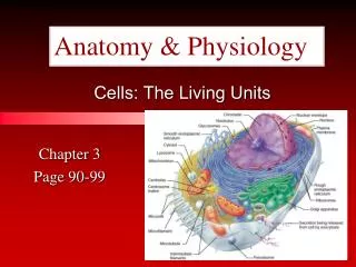
Structure of a Generalized Cell Figure 3.2
Plasma Membrane • Separates intracellular fluids from extracellular fluids • Plays a dynamic role in cellular activity • Glycocalyx is a glycoprotein area abutting the cell that provides highly specific biological markers by which cells recognize one another
Fluid Mosaic Model • Double bilayer of lipids with imbedded, dispersed proteins • Bilayer consists of phospholipids, cholesterol, and glycolipids • Glycolipids are lipids with bound carbohydrate • Phospholipids have hydrophobic and hydrophilic bipoles
Fluid Mosaic Model Figure 3.3
Functions of Membrane Proteins • Transport • Enzymatic activity • Receptors for signal transduction Figure 3.4.1
Passive Membrane Transport: Diffusion • Simple diffusion – nonpolar and lipid-soluble substances • Diffuse directly through the lipid bilayer
Passive Membrane Transport: Osmosis • Occurs when the concentration of a solvent is different on opposite sides of a membrane • Osmosis: diffusion of water across a semi-permeable membrane • Osmolarity – total concentration of solute particles in a solution • Tonicity – how a solution affects cell volume
Effects of Solutions of Varying Tonicity • Isotonic – solutions with the same solute concentration as that of the cytosol • Hypertonic – solutions having greater solute concentration than that of the cytosol • Hypotonic – solutions having lesser solute concentration than that of the cytosol
Effect of Membrane Permeability on Diffusion and Osmosis Figure 3.8b
Passive Membrane Transport: Filtration • The passage of water and solutes through a membrane by hydrostatic pressure • Pressure gradient pushes solute-containing fluid from a higher-pressure area to a lower-pressure area
Diffusion Through the Plasma Membrane Figure 3.7
Passive Membrane Transport: Diffusion • Facilitated diffusion • Transport of glucose, amino acids, and ions • Transported substances bind carrier proteins or pass through protein channels • Are integral trans-membrane proteins • Show specificity for certain polar molecules including sugars and amino acids
Process of Facilitated Diffusion • Protein binds with molecule • Shape of protein changes • Molecule moves across membrane
Facilitated Diffusion Ion CHANNELS Membrane proteins create a tunnel through which Ions can pass. http://bio.winona.edu/berg/ANIMTNS/voltgate.htm
Active Transport Transport that require energy to move molecules against the concentration gradient. Cell example: Want to put MORE glucose into mitochondria when there is already glucose in there. Image from: http://www.biologyclass.net/mitochondria.jpg
Active Transport • Two Types of Active Transport. • Pumps • Cytosis.
Pumps • Carrier proteins that change shape for molecules that are not the correct shape. • Completely changing shape requires energy.
Sodium-Potassium Pump Extracellular fluid K+ is released and Na+ sites are ready to bind Na+ again; the cycle repeats. 6 Binding of cytoplasmic Na+ to the pump protein stimulates phosphorylation by ATP. 1 Cytoplasm Phosphorylation causes the protein to change its shape. 2 Concentration gradients of K+ and Na+ The shape change expels Na+ to the outside, and extracellular K+ binds. 3 Loss of phosphate restores the original conformation of the pump protein. 5 K+ binding triggers release of the phosphate group. 4 Figure 3.10
Vesicular Transport • Transport of large particles and macromolecules across plasma membranes • Exocytosis – moves substance from the cell interior to the extracellular space • Endocytosis – enables large particles and macromolecules to enter the cell
Vesicular Transport • Transcytosis – moving substances into, across, and then out of a cell • Vesicular trafficking – moving substances from one area in the cell to another
Exocytosis Figure 3.12a
Forms of Endocytosis • Phagocytosis – cell eating • Pinocytosis – cell drinking
WHITE BLOOD CELL ENGULFING BACTERIA(Phagocytosis) http://fig.cox.miami.edu/~cmallery/255/255ion/fig14x28.jpg
INSULIN being released by pancreas cells using exocytosis http://fig.cox.miami.edu/~cmallery/255/255ion/fig14x26.jpg
Functions of Membrane Proteins • Intercellular adhesion • Cell-cell recognition • Attachment to cytoskeleton and extracellular matrix Figure 3.4.2
Plasma Membrane Surfaces • Differ in the kind and amount of lipids they contain • Glycolipids are found only in the outer membrane surface • 20% of all membrane lipid is cholesterol
Membrane Junctions • Tight junction – impermeable junction that encircles the cell • Desmosome – anchoring junction scattered along the sides of cells • Gap junction – a nexus that allows chemical substances to pass between cells
Membrane Junctions: Tight Junction Figure 3.5a
Membrane Junctions: Desmosome Figure 3.5b
Membrane Junctions: Gap Junction Figure 3.5c