Download
1 / 11
120 likes | 329 Views
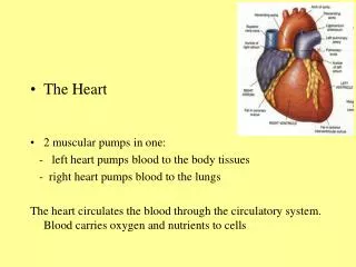
Tissues of the Heart. Sammy Meilink Gabby Avanzado Michaela Karandziff Colin Hopper. Cardiac Muscle Cells. These cells are smaller than muscle fibers They contain a single, central nucleolus Contains myofibrils and contraction – involves shortening of individual sarcomeres

E N D
Tissues of the Heart Sammy Meilink Gabby Avanzado Michaela Karandziff Colin Hopper
Cardiac Muscle Cells • These cells are smaller than muscle fibers • They contain a single, central nucleolus • Contains myofibrils and contraction – involves shortening of individual sarcomeres • Dependent on aerobic metabolism to obtain energy to continue contracting • There are reserves that store energy as glycogen and lipids
Chordae Tendineae • Blood travels from the right atrium to the right ventricle through a broad opening bounded by three flaps of fibrous tissue • Part of the right atrioventricular – also known as tricuspid • Each cusp is braced by connective tissue fibers
Chordae Tendineae http://www.google.com/imgres?q=chordae+tendineae&hl=en&safe=active&sa=X&rls=com.microsoft:en-us:IE-SearchBox&biw=1024&bih=600&tbm=isch&prmd=imvns&tbnid=Dr0sTllHS6ExFM:&imgrefurl=http://fyeahmedicine.tumblr.com/post/4089259037/white-coat-chordae-tendineae-in-the-heart&docid=z6YbsE4M9_Zs7M&imgurl=http://24.media.tumblr.com/tumblr_limhrks6cE1qaqphpo1_500.jpg&w=500&h=401&ei=DUiET9zBBIL-9QTB54TPCA&zoom=1&iact=hc&vpx=97&vpy=264&dur=2531&hovh=201&hovw=251&tx=142&ty=200&sig=113621735808706121368&page=1&tbnh=132&tbnw=169&start=0&ndsp=18&ved=1t:429,r:12,s:0,i:95
Papillary Muscle • Chordae Tendineae is connected to the papillary muscles • Cone shaped projections on the inner surface of the ventricle
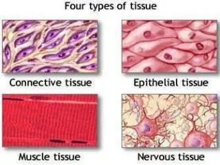
Connective Tissue • Abundant collagen and elastic fibers wrap around cardiac muscle cells and tie together adjacent cells
Three Functions • Provide support for cardiac muscle fibers, blood vessels, and nerves of myocardium • Add strength and prevent overexpansion of the heart • Help the heart return to normal shape after contractions
Layers of the Heart • Intercalated discs interlock membranes of adjacent cells by desmosomes and linked by gapped junctions • They convey the force of contraction from cell to cell • Provide movement of ions and small molecules
Layers of the Heart cont. • Epicardium covers the outer surface of the heart • Myocardium contains cardiac muscle tissue, blood vessels and nerves • Endocardium a simple squamous epithelium that is continuous
Vocabulary • Intercalated discs: Each cardiac muscle cell is in contact with several others at specialized sites • Desmosomes: The cell membranes of two cells are locked together by intercellular cement and by membrane proteins connected to a network of intermediate filaments • Intercellular cement: Is a protein-polysaccharide mixture • Myofibrils: Organized collections of myofilaments in skeletal and cardiac muscle cells • Myofilaments: Protein filament consisting of the proteins actin and myosin
Work Cited • Slide Two Pic: http://www.google.com/imgres?q=cardiac+muscle+cell&hl=en&sa=X&qscrl=1&nord=1&rlz=1T4ACEW_enUS378US379&biw=624&bih=889&tbm=isch&prmd=imvns&tbnid=FfPmAz7Acna-RM:&imgrefurl=http://histologyolm.stevegallik.org/node/146&docid=K2HQhrvAF7pWnM&imgurl=http://stevegallik.org/sites/histologyolm.stevegallik.org/images/cardiacmuscle.jpg&w=600&h=471&ei=DMCGT_GNO4my2QXfqvDlCA&zoom=1&iact=rc&dur=1&sig=116640695688943885148&page=2&tbnh=151&tbnw=192&start=12&ndsp=15&ved=1t:429,r:2,s:12,i:146&tx=113&ty=99 • Slide Four Pic: http://www.google.com/imgres?q=chordae+tendineae&hl=en&safe=active&sa=X&rls=com.microsoft:en-us:IE-SearchBox&biw=1024&bih=600&tbm=isch&prmd=imvns&tbnid=Dr0sTllHS6ExFM:&imgrefurl=http://fyeahmedicine.tumblr.com/post/4089259037/white-coat-chordae-tendineae-in-the-heart&docid=z6YbsE4M9_Zs7M&imgurl=http://24.media.tumblr.com/tumblr_limhrks6cE1qaqphpo1_500.jpg&w=500&h=401&ei=DUiET9zBBIL-9QTB54TPCA&zoom=1&iact=hc&vpx=97&vpy=264&dur=2531&hovh=201&hovw=251&tx=142&ty=200&sig=113621735808706121368&page=1&tbnh=132&tbnw=169&start=0&ndsp=18&ved=1t:429,r:12,s:0,i:95 • Slide Five Pic: http://www.google.com/imgres?q=papillary+muscle&hl=en&sa=X&qscrl=1&nord=1&rlz=1T4ACEW_enUS378US379&biw=624&bih=889&tbm=isch&prmd=imvns&tbnid=yEOe3S5LZp4fSM:&imgrefurl=http://cardiacsurgeryacademy.org/Structure-of-different-heart-Valves.html&docid=02cjwJB5f40eUM&imgurl=http://98.131.235.9/images/Anatomy-of-cardiac-valves.jpg&w=423&h=301&ei=esCGT6mjEca22gWMk9mRCQ&zoom=1&iact=rc&dur=1&sig=116640695688943885148&page=2&tbnh=164&tbnw=246&start=12&ndsp=15&ved=1t:429,r:5,s:12,i:113&tx=172&ty=74 • Slide Six Pic: http://www.google.com/imgres?q=connective+tissue+in+the+heart&hl=en&sa=X&qscrl=1&nord=1&rlz=1T4ACEW_enUS378US379&biw=624&bih=889&tbm=isch&prmd=imvns&tbnid=fN-hD8j8ObctmM:&imgrefurl=http://www.ouhsc.edu/histology/text%2520sections/cardiovascular.html&docid=-LqB6Z6kXvu4DM&imgurl=http://www.ouhsc.edu/histology/Glass%252520slides/78_07.jpg&w=400&h=314&ei=RcGGT-jxEsmw2QXsvKjnCA&zoom=1&iact=rc&dur=1&sig=116640695688943885148&page=1&tbnh=152&tbnw=194&start=0&ndsp=12&ved=1t:429,r:1,s:0,i:71&tx=65&ty=53 • Slide Eight Pic: http://www.google.com/imgres?q=intercalated+discs&hl=en&sa=X&qscrl=1&nord=1&rlz=1T4ACEW_enUS378US379&biw=624&bih=889&tbm=isch&prmd=imvns&tbnid=hwZkCLFyMOBjnM:&imgrefurl=http://neuromedia.neurobio.ucla.edu/campbell/muscle/wp.htm&docid=aGj76SZpDlm6sM&imgurl=http://neuromedia.neurobio.ucla.edu/campbell/muscle/wp_images/17_intercalated_disk.gif&w=640&h=512&ei=lcGGT7jPIeOS2QW6t7DlCA&zoom=1&iact=rc&dur=120&sig=116640695688943885148&page=1&tbnh=142&tbnw=178&start=0&ndsp=13&ved=1t:429,r:11,s:0,i:147&tx=104&ty=62 • Slide Nine Pic: http://www.google.com/imgres?q=layers+of+the+heart&hl=en&sa=X&qscrl=1&nord=1&rlz=1T4ACEW_enUS378US379&biw=624&bih=889&tbm=isch&prmd=imvns&tbnid=AEs0HCYB5VH2BM:&imgrefurl=http://www.science-art.com/image/%3Fid%3D3885%26m%3D154&docid=VQ0LiuKTJNiZzM&imgurl=http://www.science-art.com/gallery/154/154_31920092831.jpg&w=405&h=321&ei=6MGGT_WSDajO2gX6vZyJCQ&zoom=1&iact=hc&vpx=2&vpy=130&dur=537&hovh=200&hovw=252&tx=171&ty=110&sig=116640695688943885148&page=2&tbnh=148&tbnw=187&start=12&ndsp=17&ved=1t:429,r:3,s:12,i:175 • Essentials of Human Anatomy by Bartholemez