Download
1 / 47
470 likes | 587 Views
Early Brain Development in Normal and High Risk Children. John H. Gilmore, MD Department of Psychiatry The University of North Carolina. Neurodevelopmental Hypothesis of Schizophrenia. Neurodevelopmental disorder with prenatal/perinatal origins Pregnancy and birth complications (OR 2.0-4.0)

E N D
Early Brain Development in Normal and High Risk Children John H. Gilmore, MD Department of Psychiatry The University of North Carolina
Neurodevelopmental Hypothesis of Schizophrenia • Neurodevelopmental disorder with prenatal/perinatal origins • Pregnancy and birth complications (OR 2.0-4.0) • Subtle childhood neurodevelopmental abnormalities • Brain abnormalities on MRI are present at first episode
Abnormal Cortical Connectivity • Postmortem studies • reduced neuropil • decreased synaptic markers • Synaptophysin, decreased spine numbers • no overall neuron loss • Abnormal functional connectivity on fMRI
Reduced Synapses/Spines Subject with schizophrenia Matched normal control subject Glantz and Lewis, 1997 Glantz and Lewis, 2000
Synaptophysin Prefrontal Ctx Glantz et al., 2007
Schizophrenia as a neurodevelopmental disorder • Hypothesized that the structural brain abnormalities associated with schizophrenia arise during very early brain development • No direct evidence to support this hypothesis • To understand the origins of schizophrenia and other neurodevelopmental disorders, it is critical to develop methodologies to study prenatal and neonatal brain structure
Neonatal MRI: 3T high resolution, high speed scans FSE PDw 1.25 x 1.25 x 1.95mm3 FSE T2w 1.25 x 1.25 x 1.95 mm3 T1 3D MPRage 1.0 x 0.9 x 0.9 mm3 3T Siemens Allegra Scan Time: Structural MRI (T1, SpinEcho): 8min, DTI: 4min -> 12 Min tot
Neonatal MRI • 3T (Siemens Allegra head-only) • Unsedated, outpatient setting • Neonates are fed prior to scanning, swaddled, fitted with ear protection; heads fixed in a vac-fix device • A pulse oximeter monitored by a physician or research nurse • Most neonates sleep during the scan • Motion-free scans in approximately 83%
Safety Issues • Scanner is FDA approved for use in all ages • Scanner software and hardware limits specific absorption rates to safe levels based on infant weight • Phantom study with scan sequences • Mean (SD) increase 0.19±0.20 ºC • Range 0.0-0.5 ºC • (Gilmore et al., Psych Res: Neuroimaging, 132, 2004)
Study Approach • Prenatal ultrasound, neonatal MRI • Neurostructural phenotype • Enlarged lateral ventricles • Gray matter, white matter development • Two high risk groups • Genetic high risk: offspring of mothers with schizophrenia (10% develop schizophrenia) • Structural high risk: fetuses with isolated mild ventriculomegaly
Study Design • Prenatal ultrasound at 22 and 32 weeks • MRI at 2 weeks after birth • Developmental assessments at 1 and 2 years of age • Mullen Scales of Early Learning • Working Memory, Attention
Early Brain Development Studies • Recruiting to date • Mothers with schizophrenia 47 • Fetuses with mild ventriculomegaly 50 • Controls 257 • Twins 158 pairs • Bipolar 33 • Successful neonatal MRI’s to date • Mothers with schizophrenia 29 • Fetuses with mild ventriculomegaly 37 • Controls 195 • Twins 110 pairs • Bipolar 11
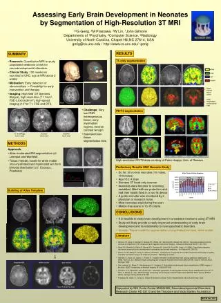
Challenges of Tissue Segmentation • Small head size • Low contrast • Bias field / intensity inhomogeneity • Motion artifacts • Ambiguous classification of white matter into myelinated and non-myelinated white matter
csf nWM gm mWM Automated Tissue Segmentation T2 T1 Prastawa M, Gilmore JH, Lin W, Gerig G Med Image Anal 2005; 9: 457-466 Early Myelination
Neonatal Brain Development Overall homogeneity of slopes: p < 0.001 Gray Matter vs. White Matter: p <0.001 Gray Matter vs. CSF: p < 0.001 Gray Matter vs. Umyelinated WM: p < 0.001
Regional Gray Matter Overall homogeneity of slopes: p < 0.001 Occipital vs. Prefrontal: p <0.001 Parietal vs. Prefrontal: p < 0.001
Regional White Matter Overall homogeneity of slopes: p = 0.12
Conclusions • Early neonatal brain development is characterized by rapid increases in gray matter compared to white matter • Regional specificity of gray matter development: posterior faster than anterior • Gender differences in ICV, gray matter volumes present at birth • Arise during prenatal brain development • Asymmetries present at birth, L>R • Adult pattern develop after birth
Isolated Mild Ventriculomegaly • Atrial width ≥ 10mm • No associated CNS abnormalities • Up to 0.7% of pregnancies • Associated with older maternal age, lower gestational age at birth, and maternal infection • Gilmore et al., 1998; Dommergues et al., 1996 • Outcome • 33% have developmental delays (Bloom et al., 1997) • Autism, ADHD, learning disorders (Gilmore et al., 2001)
MVM study • 34 children with isolated MVM • 34 age and gender matched controls • Children in the MVM group had significantly larger prenatal maximum lateral ventricle width • 1.15 ± 0.03 vs. 0.59 ±0.03; p < 0.0001
Lateral Ventricles • Maximum lateral ventricle width in controls and MVM cases (n= 34/ group; p < 0.0001) • Neonates with prenatal MVM have significantly larger lateral ventricle volumes than • matched controls (n= 34/ group; p < 0.0001).
Prenatal/Neonatal Relationship There was a significant correlation between the prenatal maximum lateral ventricle width on ultrasound and neonatal lateral ventricle volume on MRI for both the normal control (Pearson r = 0.3563; p = 0.0386) and the MVM groups (Pearson r = 0.7482, p < 0.0001)
Gray and White Matter Volume There is a significant difference in the relationship between ICV and cortical gray matter volume in MVM cases compared to controls (homogeneity of slope F=13.15 (1,31); p=0.0010) There is a significant difference in the relationship between ICV and cortical white matter volume in MVM cases compared to controls (homogeneity of slope F= 7.04 (1,31); p=0.0125)
General Principles • Mean Diffusivity decreases with age • Fractional Anisotropy increases with age • Mean Diffusivity a more sensitive marker of myelination in neonates
MVM Conclusions • Prenatal enlargement of the lateral ventricle detected by ultrasound is associated with significant enlargement of the lateral ventricles after birth • Increased gray matter volumes • Reduced white matter volumes, and delayed or abnormal maturation of DTI properties in the splenium of the corpus callosum • It is suggested that prenatal ventricle volume may be an early structural marker of subsequent dysmaturation of the cerebral cortex after birth
Offspring of Mothers with Schizophrenia • Neonatal MRIs on 19 high risk children and 19 matched controls • Mothers with schizophrenia, schizoaffective DO • Controls without psychiatric illness • Matched on gender, maternal age, gestational age at birth, ethnicity • 9 males and 10 females • mean gestational age at MRI 42.7 ± 3.0 weeks
Neonatal brain structure in high risk children p = 0.0325 p = 0.083 • High risk children had approximately 2.6% less total gray matter (p = 0.077)
Conclusions • Early results indicates that the offspring of mothers with schizophrenia have reduced cortical gray matter volumes in the rapidly developing occipital region • May reflect genetically mediated impairment of cortical synapse development that would be most apparent in the rapidly growing cortical region • There is a suggestion of altered white matter development • No difference in lateral ventricle volumes • Lateral ventricle volumes increase rapidly in the first year of life – the enlargement may arise after birth • These results focus the time-frame of candidate neurodevelopmental processes that contribute to risk for schizophrenia • Limitations • Medications during pregnancy • Mothers with schizophrenia have high rates of prenatal/perinatal complications
Early Brain Development in 1 and 2 year Olds • Singleton Controls • 59 one year olds (68% success rate) • 44 two year olds (60% success rate) • Twins • 51 pairs at age 1 (90% success rate) • 37 pairs at age 2 (76% success rate)
T1w T2w 2 weeks 1 year 2 years Subject with follow-up scans
Brain development birth to age 2 TBV grows 101% in first year, 15% in second year 2-4 weeks: 36% of adult volume; 72% at 1 year and 83% at 2 years
Brain development birth to age 2 Cortical GM: 149% in the first year; 14% in the second year Cortical WM:
Future Directions • Collecting DNA to study gene-brain structure relationships in early childhood • Developmental assessments at age one and two years to study structure-function relationships • Develop age specific head coils to improve resolution and contrast (W. Lin) • Resting State Networks (W. Lin) • Apply to other high risk groups
Acknowledgements • MRI Acquisition • Weili Lin PhD, Keith Smith MD, Kathy Wilber • Image Analysis • Guido Gerig PhD, Martin Styner, PhD, Sampath Vetsa, Marcel Prastawa, Isabelle Corouge, Sylvain Gouttard, Christopher Looney • Dinggang Shen, PhD • Statistics/Data Management • Robert Hamer PhD, Chaeryon Kang, Abby Scheer MA • Study Coordinator • Dianne Evans MA