Download
1 / 12
120 likes | 213 Views
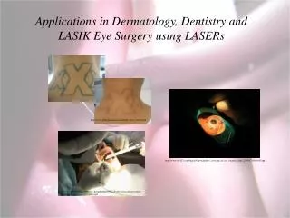
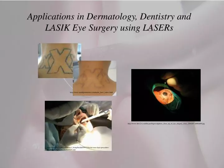
Applications in Dermatology, Dentistry and LASIK Eye Surgery using LASERs. http:// www.medispainstitute.com/menu_laser_tattoo.html. http://www.life123.com/bm.pix/bigstockphoto_close_up_of_eye_surgery_catar_2264267.s600x600.jpg.

E N D
Applications in Dermatology, Dentistry and LASIK Eye Surgery using LASERs http://www.medispainstitute.com/menu_laser_tattoo.html http://www.life123.com/bm.pix/bigstockphoto_close_up_of_eye_surgery_catar_2264267.s600x600.jpg http://www.ny1.com/content/ny1_living/health/89972/doctor-uses-laser-procedure-to-eliminate-gum-disease/Default.aspx
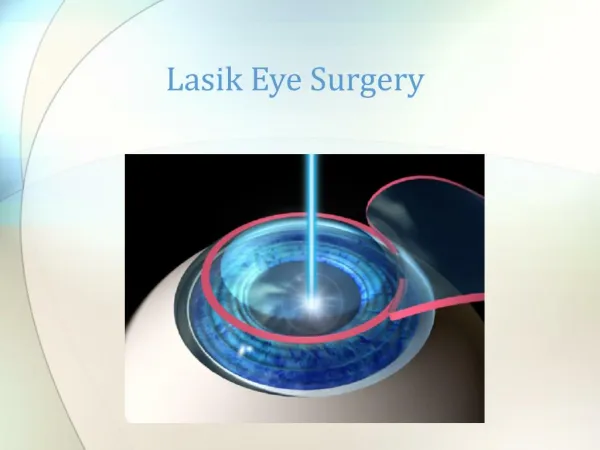
LASIK Eye Surgery • LASIK is an acronym of Laser Assisted In-Situ Keratomileusis or the subsequent use of an laser (usually an eximer laser) to reshape and flatten the cornea after an incision has been made in the cornea by using a microkeratome (a thin knife) or a laser keratome. • The Epithelium is incised and folded back revealing the Stroma. • The stroma is laser vaporized to reshape the cornea and the epithelium is then replaced. 1. Epithelium (cornea) 2. Stroma (cornea) 3. Descemet's membrane and endothelium (cornea) 4. Anterior chamber 5. Iris 6. Lens 7. ciliary body 8. sclera http://www.missionforvisionusa.org/anatomy/uploaded_images/GrosASlabMfV-702936.jpg
LASIK Eye Surgery - What for? • In a nutshell, the cornea aids in the focusing of light to create an image on the retina by means of refraction. • Often, the shape of the cornea and the eye are not perfect and the image on the retina is out-of-focus. • There are three primary types of refractive errors (or imperfections in the focusing power of the eye.) • Myopia – or nearsightedness • Persons with myopia, or nearsightedness, have more difficulty seeing distant objects as clearly as near objects. • Hyperopia - or farsightedness • Persons with farsightedness have more difficulty seeing near objects as clearly as distant objects. • Astigmatism – which is a distortion of the image on the retina caused by irregularities in the cornea or lens of the eye (usually due to the cornea not being spherical, but oval in shape.)
LASIK Eye Surgery - What for? • Nearsightedness, or myopic vision, the image forms in front of the retina while in the case of farsightedness, or hyperopic vision, the image forms behind the retina. • Combinations of myopia and astigmatism or hyperopia and astigmatism are common and can be corrected with glasses or contact lenses that are designed to compensate for the eye's imperfections. • Surgical procedures aimed at improving the focusing power of the eye are called refractive surgeries. • In a LASIK surgery, precise and controlled removal of • corneal tissue by a special laser reshapes the cornea • changing its focusing power and aids in the eyes • ability to focus the light. • For myopia, microthin layers of cornea are eliminated • to flatten its shape. • For hyperopia, a doughnut-shaped hole is made to • create a more conical shape. Jay Newman: Physics of the Life Sciences, Springer, 2009
LASIK Eye Surgery - Anatomy of the eye • The cornea is the transparent, dome-shaped window covering the front of the eye. It is a powerful refracting surface, providing 2/3 of the eye's focusing power. Like the crystal on a watch, it gives us a clear window to look through. • There are no blood vessels in the cornea, and it is normally clear with a shiny surface. The cornea is extremely sensitive - there are more nerve endings in the cornea than anywhere else in the body. • The adult cornea is only about ½ millimeter thick. http://www.stlukeseye.com/anatomy/Cornea.asp http://www.shorelinevision.com/visiondata/db/UPLOADEDIMAGES/cornea2-20071103-164958.jpg
LASIK Eye Surgery - Anatomy of the eye • The crystalline lens is located just behind the iris. Its purpose is to focus light onto the retina. The nucleus, the innermost part of the lens, is surrounded by softer material called the cortex. The lens is encased in a capsular-like bag and suspended within the eye by tiny delicate fibers called zonules. • In young people, the lens changes shape to adjust for close or distance vision. This is called accommodation. With age, the lens gradually hardens, diminishing the ability to accommodate. http://www.stlukeseye.com/anatomy/Lens.asp An actual photograph of a human eye that has been bisected in the coronal plane to show the view of the anterior segment from a posterior perspective (as though you are looking from the retina). The crystalline lens is suspended by delicate fibers called the zonule. The ciliary body (CB) is composed of about 72 processes that make up the pars plicata and a flat area called the pars plana. The oraserrata (ora) is the place where the retina joins the ciliary body. http://www.missionforvisionusa.org/anatomy/2005/10/cornea-iris-lens-anterior-segment-of.html
LASIK Eye Surgery - Anatomy of the eye • The colored part of the eye is called the iris and the iris controls light levels inside the eye similar to the aperture on a camera. The round opening in the center of the iris is called the pupil. The iris is embedded with tiny muscles that dilate (widen) and constrict (narrow) the pupil size. • The sphincter muscle lies around the very edge of the pupil. In bright light, the sphincter contracts, causing the pupil to constrict. The dilator muscle runs radially through the iris, like spokes on a wheel. This muscle dilates the eye in dim lighting. • The iris is flat and divides the front of the eye (anterior chamber) from the back of the eye (posterior chamber). Its color comes from microscopic pigment cells called melanin. The color, texture, and patterns of each person's iris are as unique as a fingerprint. http://www.stlukeseye.com/anatomy/Iris.asp http://www.cl.cam.ac.uk/~jgd1000/sampleiris.jpg
LASIK Eye Surgery - Anatomy of the eye • The retina is a multi-layered sensory tissue that lines the back of the eye. It contains millions of photoreceptors that capture light rays and converts the light into electrical impulses. These impulses travel along the optic nerve to the brain where they are turned into images. • There are two types of photoreceptors in the retina: rods and cones. • The retina contains approximately 6 million cones and they are contained in the macula, the portion of the retina responsible for central vision. They are most densely packed within the fovea, the very center portion of the macula. Cones function best in bright light and allow us to appreciate color. • There are approximately 125 million rods. They are spread throughout the peripheral retina and function best in dim lighting. The rods are responsible for peripheral and night vision. http://www.stlukeseye.com/anatomy/Retina.asp
LASIK Eye Surgery - Anatomy of the eye: a closer look at rods & cones Suzanne Amadore Kane: The Physics of Modern Medicine Jay Newman: Physics of the Life Sciences, Springer, 2009 Clockwise from the upper left: An sketch of the retina showing its structure, The pigment epithelium showing the rods and cones. Two SEM pictures of the cone cells. Jay Newman: Physics of the Life Sciences, Springer, 2009 Jay Newman: Physics of the Life Sciences, Springer, 2009
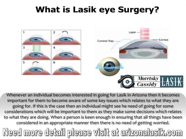
LASIK Eye Surgery - The procedure • A laser keratome or a microkeratome is used to cut a flap in the epithelium (cornea.) A hinge is left at one end of this epithelium layer. The flap is folded back revealing the stroma, and laser pulses are used to vaporize a portion of the stroma and the epithelium layer is then replaced. • In cases of myopia, microthin layers of cornea are eliminated to flatten its shape. • For hyperopia, a doughnut-shaped hole is made to create a more conical shape. • An Eximer laser – A low power (1 – 20W) UV (~ 250nm) laser which is absorbed by the cornea and not transmitted into the eye. http://www.stlukeseye.com/anatomy/Cornea.asp http://www.shorelinevision.com/visiondata/db/UPLOADEDIMAGES/cornea2-20071103-164958.jpg http://www.imhoffeye.com/lasik.htm
LASIK Eye Surgery - LASIK eye surgery video It it worth it? Benefits: improved vision fast recovery time lower risk of infections than other eye surgeries less pain than regular corneal surgery no more glasses or contacts (hmm?) recreational and job opportunities Drawbacks: light sensitivity some pain swelling of the cornea wrinkling of the epithelium flap poor night vision – halos and glare no actual measure of how much tissue to remove can’t stop the aging process – might still need reading glasses (presbyopia)
Homework for Friday, Read Kane Chapter 4, sections 4.1 – 4.5 Wolbarst Chapter 11, sections 11.1 – 11.6