Download
1 / 81
990 likes | 2.86k Views
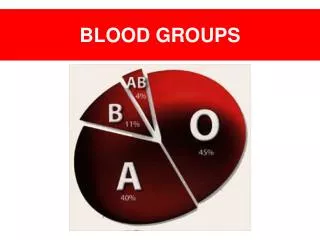
Other Blood Groups Lewis, Kell, Duffy, Kidd, Ii, MNSs & P. Introduction. Over 500 blood group antigens “High incidence”, “public” or “high frequency” antigens are those present on almost every person’s red blood cells

E N D
Other Blood Groups Lewis, Kell, Duffy, Kidd, Ii, MNSs & P
Introduction Over 500 blood group antigens “High incidence”, “public” or “high frequency” antigens are those present on almost every person’s red blood cells “Low incidence”, “private” or “low frequency” antigens are present on very, very few individuals red blood cells
Introduction Each known antigen initially identified through the detection of its specific antibody in the serum. Knowledge of serologic behavior and characteristics of blood group antibodies is CRITICAL for identification
Introduction Essential when evaluating antibody screen and panel studies. Considerations given to: Phase of reactivity Antibody class involved Ability to cause HDFN and HTR
Major Blood Group Systems Lewis I P MNSs Kell Kidd Duffy
Lewis System Major antigens Lea and Leb , other antigens include Lec, Led and Lex Antigens ARE NOT intrinsic to RBCs but are absorbed from the plasma and inserted into RBC membrane.
Lewis System Antigenic Development Genetic control reside in single gene “Le” Amorph le, if homozygous will not have Lewis antigens Lea formed first, then modified to form Leb which is adsorbed preferentially over Lea Lewis phenotype of RBC can be changed by incubating with plasma containing Lea or Leb glycoplipid.
Lewis System Lewis antigens in infants Antigens absent or extremely weak at birth Expression of Leb gradual Birth Le (a-b-) 2 months Le(a+b-) 12 to 18 months Le(a+b+) 2 to 3 years Le (a-b+)
Lewis System Lewis antigens and pregnancy Antigen strength may decline dramatically Transiently Le (a-b-) may produce Lewis antibodies during pregnancy Antigens return after delivery and antibodies disappear
Lewis System Interaction of Le, Se and H Genes lele will not have Lewis antigens, but if Se present will have A, B and H in secrections Genotype se/se and have one Lewis antigen will have Lea in their secretions but no A, B or H.
Lewis System Lewis Antibodies Almost always IgM, react strongly at RT, may cause ABO discrepancy if reverse cells have Lewis antigen. Occur almost exclusively in Le (a-b-) and production of anti-Lea AND –Leb not unusual Anti-Lea frequently encountered, anti-Leb rarely encountered.
Lewis System Lewis Antibodies Although most react at RT reactivity may be seen at 37C, but is weaker and may be weakly reactive at AHG Can bind complement and cause IN-VITRO hemolysis, most often with enzyme treated cells Because antibodies are IgM and antigens are poorly developed at birth antibodies NOT implicated in HDFN.
Lewis System Lewis antibodies Can be neutralized in-vitro by additions of Lewis Substance Le antigens are present in secretions Add to serum with Lewis antibodies and the antibodies will be bound to the soluble Lewis antigens Useful when multiple antibodies are present and 1 is a Lewis, eliminates the activity of the antibody
Lewis Antibodies • Anti-Le a, Anti-Le b, Anti-Lex • Most react at room temperature or below - • Often fix complement • Some in vitro hemolysis • Le a may cause HTR
Lewis Antibodies • Anti-Le a • Found in Lea-b- secretors • best room temperature or below • Often fix complement • Some in vitro hemolysis • Le a may cause HTR
Lewis Antibodies • Anti-Le b • Often found with Anti-Lea • Most react at room temperature or below • Two types - Anti-LebH and Anti-LebL • Rare cause of HTR
Lewis Antibodies • Anti-Lex • Most react at room temperature or below - • Reacts with both Lea and Leb as a single antibody
Lewis Antibodies • Special Problems in the Blood Bank • Lewis antigens may be weaker during pregnancy and women produce antibodies • Can neutralize Lewis antibodies with Lewis plasma • Pregnant woman with room temperature antibodies, neutralize with Lewis antigen when testing for HDN antibodies
Lewis System Transfusion Practice Transfused RBCs will acquire the Lewis phenotype of the recipient within a few days Lewis antibodies in patient will be neutralized by Lewis substance in donor plasma Lewis antibodies rarely cause in-vivo hemolysis It is not necessary to phenotype donors for Lewis antigens prior to transfusion, give crossmatch compatible
Background information • The Kell blood group system was discovered in 1946. • Number of Kell antigens: > 20 • These antigens are the third most potent, after those of the ABO and Rh blood groups, at triggering an immune reaction.
Molecular information • The KEL gene is found on chromosome 7 • The KEL gene is highly polymorphic, with different alleles at this locus encoding the 25 antigens that define the Kell blood group. • The Kell protein is a polypeptide chain of 732 amino acids in length that becomes glycosylated at five different sites. It makes a single pass through the RBC membrane.
Kell Blood Group System • XK gene produces Kx substance, which is a precursor of of Kell Ags • Kel genes convert Kx substance into the Kell Ags on RBCs • K (Kell) & k (cellano) are produced by allelic genes, this results into 3 phenotypes: • K+k- (genotype KK) • K+k+ (genotype Kk) • K-k+ (genotype kk) • Other allelic genes include: Kpa/Kpb, Jsa/Jsb
XK Gene (Chromosome X) KEL Gene RBC Kell system glycoprotein: Kell Ag’s reside here. Kx
Kx Substance • Kx substance is present on RBCs & WBCs • Kell genes convert Kx substance into the Kell Ags on RBCs • Kell genes do not convert Kx on WBCs
McLeod Phenotype • Absence of Kx proteins in RBCs membrane lead to McLeod Phenotype • This absence cause: • abnormal RBCs shape (acanthocytes) • & reduced in-vivo survival
Chronic Granulomatous Disease • Absence of Kx proteins in WBCs cause CGD • Leukocytes are able to phagocytose but not to kill bacteria • Patients with CGD have recurrent bacterial infections • Patients who lack Kx on RBCs & WBCs have both Mcleod and CGD
Kell Antibodies • K- individuals produce anti-K when exposed to K+ cells • Frequency of K+ is low (9%), easy to find blood • On the other hand frequency of k is 99.9% • k- individuals produce anti-k when exposed to k+ cells • Difficult to find blood
Duffy Blood Group System • The Duffy blood group was discovered in 1950. • The Duffy glycoprotein is encoded by the FY gene, found on chromosome 1 , of which there are two main alleles, FYA and FYB. They are codominant. • The Duffy gene codes for a glycoprotein also found in other tissues: brain, kidney, spleen, heart and lung. • The Duffy glycoprotein is a transmembrane protein • Five alleles at Duffy locus, the most important: Fya, Fyb & Fy (Silent Allele) • Fya is more immunogenic than Fyb
Duffy Antigens • Phenotype Frequencies
Different genes • Fy(a-b-) blacks do not produce anti-Fya oranti-Fyb following transfusion with Fy(a+) or Fy(b+) blood • Fy(a-b-) Caucasians become sensitizedfollowing transfusion with Fy(a+) or Fy(b+) blood • This suggest that Fy(a-b-) phenotype arises from different genes in the two populations
Duffy Antigens • Fya, Fyb antigens are Destroyed by enzymes • Abs DO NOT agglutinate enzyme treated cells • Moderately immunogenic • Fya is more immunogenic than Fyb
Duffy Antibodies • IgG antibodies and can activate complement • Anti- Fya is more frequently encountered • Anti- Fyb is more frequently found in patients produced multiple alloantibodies
Duffy and Malaria • Black people with the Duffy phenotype of Fy(a–b–) appear to have resistance to Plasmodium vivax & Plasmodium knowlesi causative agents of Malaria. • Duffy antigens appear to be a receptor for the P. vivax organism and when the antigen is not present on the red blood cell membrane P. vivax is unable to access the red blood cell • Some area’s of West Africa are 100% Fy(a–b–). • Plasmodium falciparum binds to RBCs at integral glycophorin A & B
Kidd Blood Group System • The Kidd blood group was discovered in 1950. • The Kidd gene is located on chromosome 18 • Three alleles: Jka, Jkb, Jk • Codominant Inheritance • Jkis a silent allele (amorph) • The Kidd protein is an integral protein of the RBC membrane.