Download
1 / 160
1.6k likes | 1.93k Views
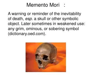
MEMENTO MORI. MAXILLO-FACIAL TRAUMA. R.Drummond October 24, 2002 preceptor: Carol Holmen. Overview. General approach to facial trauma Epidemiology anatomy diagnostic imaging specific conditions diagnosis of facial trauma as a presentation of abuse Conclusions. General Comments.

E N D
MAXILLO-FACIAL TRAUMA R.Drummond October 24, 2002 preceptor: Carol Holmen
Overview • General approach to facial trauma • Epidemiology • anatomy • diagnostic imaging • specific conditions • diagnosis of facial trauma as a presentation of abuse • Conclusions
General Comments • Injuries to the face devastating to patient • physical, emotional, occupational, sequelae • Two presentations simple, isolated injuries clinically stable vs. Manifestation of severe trauma • 25% of maxillofacial trauma involves litigation • most injuries can be picked up on thorough clinical assessment • Our role is usually to diagnose not treat • Overlap of specialists ENT, OPHTH,PLASICS, NEUROSURGERY, DENTISTRY
Question 1: The single most valuable xray of the mid-face is: • 1)Water’s view • 2)Lateral view • 3)Caldwell view • 4)Towne’s view
Question2 : Most associated injuries in cases of maxillofacial trauma are to the: • 1)brain • 2)cervical spine • 3)chest • 4)abdomen
Question 3: Open bite may be secondary to all except: • 1)LeFort Fracture • 2)tripod fracture • 3)mandibular fracture • 4)NEO fracture
Question 4: All of the following are true about children with maxillo facial trauma except • 1)greater risk of lower cervical spine injury • 2)intracrainial injury is higher • 3)mid-face fracture higher as child grows • 4)non-accidental trauma should be considered
Triage scenario • Two vehicle head on collision, driver and front seat passenger in one vehicle, • single driver in second vehicle • cars each going 30 m.p.h. • all were unrestrained • all brought to ED by EMS • all on spinal boards
Patient 1 • 5 year old child passenger of car • windshield fractured in target pattern • No LOC • Large Laceration across forehead , boggy swelling of skin, moderate “watery” epistaxis • HR 140 BP 90/45 RR 34 (crying) sats 100% • GCS 15
Patient 2 • 26 year old woman, was driver of the car • face hit steering wheel... No L.O.C. • Badly injured face, no other obvious injuries • gasping “I have to sit up I can’t breathe” • vitals HR 120 BP 90 /40 RR 36 Sats 89 on 10litres GCS 14 • primary survey gurgling resps with considerable blood in mouth gaping wounds across forehead jaw is mangled with evident deformity
Patient 3 • 18 year old driver of other vehicle works as a miniaturist painter, lost his bottle-bottom spectacles at scene of accident • hit driver’s side window • No L.O.C. • HR 100, BP 120/75 RR 24 sats 98% • GCS 15 • badly lacerated L face with deformity tender over zygoma diplopia numbness over cheek positive Marcus Gunn
Force of Gravity Necessary to Injure Face • Nasal Bones 30 x gravity • Zygoma 50 x gravity • Angle of Mandible 70 x gravity • Frontal Globellar region 80 x gravity • Midline Maxilla 100 x gravity • Supraorbital rim 200 x gravity
Basic Epidemiology • Most common causes: • MVA’s, falls, assault • community: nose and mandible • :MVA’s and Sports • urban: midface, zygoma • penetrating and assault • more than 60% have associated • other injuries
Epidemiology of MaxilloFacial Injuries at Trauma Hospitals in Ontario, Canada between 1992 and 1997The Journal Of Trauma, September 2000... Hogg et al • Ontario Trauma Registry new database • 15 -22 % of trauma patients severe maxillofacial injuries • 2,969 patients in 12 trauma centers • male: female 3:1 • most common cause mva’s • 26% positive BAC • understanding causes severity temporal distribution effective treatment and prevention
Long Term Physical Impairment and Functional Outcomes after Complex Facial FracturesPlastic and Reconstructive Surgery, August 2001 Girotto, MacKenzie et al • Retrospective cohort study of adults 18 - 25 • 265 pts with LeFort fractures compared to 242 pts with severe general injury • followed with several tools to assess health and well being • (General Health Questionnaire, Body Satisfaction Scale, Social Avoidance and Distress Scale) • hypothesis early intervention at tertiary care trauma center better results • complex facial fractures represent subset of trauma with more longterm complications
Obvious sequelae: • Diplopia 56% Zygomatic fractures • 23% LeFort fractures • 20 -31% midface fractures difficulties mastication • 35% Anasomia in LeFort Fractures • Epiphora midface fractures 25- 45 % • facial numbness 32 -35 % • 55% of facial fractures returned to work at one year compared to 70% less severe facial fractures other general injuries
“An appreciation of the long term physical and psychological sequelae of injury is essential for evaluating current treatment plans and to assist in providing appropriate counseling or referral to other healthcare professionals”
Triage and immediate management • Airway management first and major priority • be prepared for surgical airway • clear cervical spine then let patient adopt most comfortable position • caution re nasal tracheal intubation • if RSI prep for cricothyroidectomy • awake intubation • ketamine a good drug • tongue often obstructs
Shock and Hemorrhage • Maxillofacial Trauma seldom cause of shock • 60% association other injuries • If shock check for other sources • with severe facial smashes • reduce fracture plates • severe epistaxis hard to control : Foley
All patients with significant facial injuries must be presumed to have cervical spine injury until proved otherwise
History • Mechanism of injury • blunt vs. Penetrating • L.O.C.? • questions: • Do you see double? • Are there areas of numbness on your face? • Does your bite feel normal? • Which areas on your face hurt? • Does it hurt when you open your mouth and where? • Consider abuse
Physical Exam • Inside Out and bottom up • bird’s eye view and worm’s eye view • Gestalt • 90% of all facial fractures can be picked up or suspected by careful palpation • careful ocular exam visual acuity fields • subconjunctival hemorrhage • Pinpoint exam, Marcus Gunn exam • raccoon eyes, battle sign • halo test • intranasal palpation test
Allergies • Tetanus status
Anatomy • Vertical buttresses: nasal, frontal, and zygomatic maxillary give vertical stability • zygomatic temporal buttresses horizontal support
Three Zones of Facial Anatomy • UPPER: Superior Orbit and above Frontal Bone • MIDDLE: Superior Orbital rim to occlusal surface • Orbits, Nasal bones, Zygoma, Maxilla • LOWER: mandible, teeth • clinical exam should guide and direct radiological exam
Diagnostic Imaging • Standard Four Views • Waters • Caldwell • Lateral • Submentovertex • Occlusal views • Panorex
Waters View • Most valuable • prone.... Clear c-spine • draw four lines should be parallel and smooth
Caldwell View • Supplements Waters view • superior orbital rim • sinuses • orbital region • can see teardrop sign • open bomb bay door sign
Lateral View • Frontal Sinus • maxillary sinus • occasionally pterygoid plate
Submentovertex view • “Jughandle” view • Main value is to see zygomatic arch
X-rays good screening test to guide which CT scan to order and level • Ctscan most useful to grade injury and plan surgery • most useful for orbital and maxillary fractures • blowout fractures in particular • axial and coronal • can do 3-D reconstruction