Download

1 / 65
1.08k likes | 3.36k Views
Congenital Anomalies of the Kidney and Urinary Tract (CAKUT). Congenital anomalies of the kidney and urinary tract (CAKUT) constitute approximately 20 to 30 % of all anomalies identified in the prenatal period.

E N D
Congenital Anomalies of the Kidney and Urinary Tract (CAKUT)
Congenital anomalies of the kidney and urinary tract (CAKUT) constitute approximately 20 to 30 % of all anomalies identified in the prenatal period. • Defects can be bilateral or unilateral, and different defects often coexist in an individual child. • The overall rate of CAKUT in live and stillborn infants is 0.3 to 1.6 per 1000 .
The incidence is higher in women with a family history of CAKUT. • Of all antenatal renal anomalies, the most frequent abnormality is hydronephrosis, (ie, upper urinary tract dilatation). • Renal malformations are associated with non-renal congenital anomalies in about 30 % of cases
1-Renal hypoplasia : • A lower number of structurally normal nephrons, is a distinct entity separate from renal dysplasia • Unknown causes
The clinical diagnosis of renal hypoplasia is suggested when all of the following criteria are met : * Reduction of renal size by 2 standard deviations for the mean size by age * Exclusion of renal scarring by 99mTc–dimercaptosuccinic acid (DMSA) radionuclide scan * In cases of unilateral renal hypoplasia, compensatory hypertrophy of the contralateral kidney
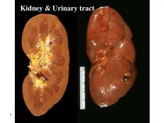
2-Renal dysplasia: • Renal dysplasia is characterized by the presence of malformed kidney tissue elements • Dysplastic kidneys are variable in size but most are smaller than normal. Size is often determined by the presence or absence of cysts. • Renal dysplasia may be unilateral or bilateral
Renal dysplasia may be discovered during routine antenatal screening or postnatally when renal ultrasonography is performed in a dysmorphic infant. • Bilateral dysplasia is likely to be diagnosed earlier than unilateral dysplasia especially if oligohydramnios is present.
Infants with bilateral dysplasia may have impaired renal function at birth and subsequent progressive renal failure may occur. • Associated urological findings include abnormalities of the renal pelvis and calyces (congenital hydronephrosis) and ureters (duplicating collecting system), megaureter, ureteralstenosis, and vesicoureteral reflux (VUR).
Because of the frequent association of renal dysplasia with a collecting system anomaly, voiding cystourethrography should be considered in all patients with renal dysplasia. • The prognosis of renal dysplasia depends on whether there is unilateral versus bilateral disease. In general, the long-term outcome of unilateral renal dysplasia is excellent, particularly if there is a normal contralateral kidney.
3-Multicystic dysplasia : • Multicystic dysplastic kidney (MCDK) is a nonfunctioning dysplastic kidney with multiple cysts, which is thought to arise from an alteration in renal parenchymal differentiation. MCDK consists of a nonreniform mass of cysts and connective tissue, and is most commonly detected by routine antenatal screening.
4-Renal agenesis: • Renal agenesis is defined as congenital absence of renal parenchymal tissue and results from major disruption of metanephric development at an early stage. • Unilateral RA accounts for 5 percent of renal malformations . • The incidence of renal agenesis is approximately 1 per 2900 births
Multiple factors are thought to be implicated in the pathogenesis of renal agenesis including mutations in genes important in renal development, and teratogenic and environmental agents (eg, retinoic acid and cocaine exposure)
Other urological abnormalities have been reported in up to 33 to 65 percent of unilateral cases • Vesicoureteral reflux (VUR) is the most commonly identified urological abnormality, • Nonrenal associated anomalies include cardiac anomalies (most commonly septal anomalies), genital tract, and gastrointestinal, respiratory, and skeletal malformations
5-Genetic cystic diseases : • Genetic cystic renal diseases are disorders of terminal epithelial differentiation A-Autosomal recessive polycystic kidney disease (ARPKD): • It is caused by mutations in the PKHD1 gene, which codes for fibrocystin. • ARPKD is characterized by multiple microscopic cysts, principally involving the distal collecting ducts Of both kidneys • Kidneys are usually greatly enlarged and contain small cysts; renal failure is common in childhood.
The liver is enlarged and has periportal fibrosis and scattered cysts. • Fibrosis produces portal hypertension by age 5 to 10 yr. • Disease severity and progression vary. Severe disease may manifest prenatally or soon after birth or in early childhood with renal-related symptoms; less severely affected patients present in late childhood or adolescence with hepatic-related symptoms.
Severely affected neonates commonly have pulmonary hypoplasia secondary to the in utero effects of renal dysfunction and oligohydramnios. • If the patient presents in adolescence, nephromegaly is less marked, renal insufficiency may be mild to moderate, and the major symptoms are those related to portal hypertension.
Diagnosis may be difficult, especially without a family history. Ultrasonography may demonstrate renal or hepatic cysts; definitive diagnosis may require biopsy. • Ultrasonography in late pregnancy usually allows presumptive in utero diagnosis. • Clinical manifestations include oligohydramnios, pulmonary hypoplasia, hypertension, congestive cardiac failure, liver disease, and renal failure. • The perinatal prognosis depends on the pulmonary status.
B-Autosomal dominant polycystic kidney disease (ADPKD) • ADPKD is characterized by bilateral renal enlargement secondary to multiple cysts. • It is caused by mutations in either PKD1 (85 percent of patients) or PKD2 genes (15 percent) • There is a greater variability in clinical manifestations of ADPKD with most patients having significant clinical findings only in adulthood.
There are a subset of children who have an early onset of disease (in utero or in the first year of life) with symptoms similar to those with ARPKD. • These include gross or microscopic hematuria, hypertension, proteinuria, cyst infection, and renal insufficiency
RENAL ECTOPY: • Renal ectopy occurs when the kidney does not normally ascend to the retroperitoneal renal fossa (level of the second lumbar vertebra). • Simple congenital ectopy refers to a kidney that lies on the correct side of the body but lies in an abnormal position.
Crossed renal ectopia • Different forms of crossed renal ectopia 1- Fused: Ectopic kidney moves across the midline and fuses to the lower pole of the normally positioned contralateral kidney. 2-Nonfused: Ectopic kidney moves across the midline without fusion and positioned at the rim of the pelvis (pelvic kidney). 3-Bilateral: Both kidneys are ectopic and cross the midline with the ureters maintaining their normal bladder insertion.
RENAL FUSION: • Renal fusion occurs when a portion of one kidney is fused to the other. • The most common fusion anomaly is the horseshoe kidney, which involves abnormal migration of both kidneys (ectopy), resulting in fusion. • This differs from crossed fused renal ectopy, which usually involves abnormal movement of only one kidney across the midline with fusion of the contralateralnoncrossing kidney.
Horseshoe kidney can be a feature of many syndromes including genetic disorders such as Turner syndrome, Trisomy 13, 18 and 2 • Patients with a horseshoe kidney appear to have an increased risk for Wilms tumor.
Most patients with an ectopic or fused kidney(s) are asymptomatic and are diagnosed coincidentally, often by antenatal ultrasonography. • In patients diagnosed symptomatically with either anomaly, symptoms at presentation are generally related to associated complications including urinary tract infection (with or without VUR), obstruction, and renal calculi.
Physical findings characteristic of bladder exstrophy in both boys and girls include: * Open bladder plate * Low set umbilicus * Diastasis of the symphysis pubis * Anteriorly displaced anus * Inguinal hernia
Urinary tract infection (UTI) is a leading cause of serious bacterial illness in febrile infants • Throughout childhood the cumulative incidence is approximately 10% in girls and 3% in boys. • Urinary infection usually is ascending, with inoculation of fecally derived organisms from the urethra and peri-urethral tissues into the bladder
The most prevalent pathogens in several recent pediatric studies were Escherichia coli (54%-67%) Klebsiella (6%-17%) Proteus (5%- 12%) Enterococcus (3%-9%) Pseudomonas (2%-6%) • Among patients with urinary tract anomalies or impaired immune systems, less virulent organisms, such as Staphepi, H influenzae, and group B Strept, may be responsible.
The hematogenous route of infection is far less common with generally different causal organisms, such as Staphaureus, Candida, and Salmonella; Pseudomonas aeruginosa and Proteus can infect by either route.
CLINICAL PRESENTATION • Young infants often present with fever alone (≥38°C); irritability, vomiting, lethargy, or poor feeding variably may be present. • For those younger than 3 months there is an increased risk of bacteremia and a greater possibility of undiagnosed congenital urologic malformations. • Older children generally have more explicit symptoms of bladder inflammation and/or flank pain.
For infants, any of the following increased the positive likelihood ratio of UTI to 2 or more: history of prior UTI, fever of more than 24 hours’ duration or higher than 40°C, absence of circumcision in males, and suprapubic tenderness. • Combinations of these findings amplified probability. • For verbal children, the following symptoms were most reliable: abdominal pain with fever higher than 38°C, back pain, new-onset urinary incontinence, dysuria, and frequency.
DIAGNOSIS OF UTI : • Specimen Collection :A non contaminated urine sample is fundamental. • For infants and non–toilet-trained children, the most accurate method of collection is suprapubic bladder aspiration, however, it rarely is practical. • Urethral catheterization or spontaneously voided clean midstream samples (usually obtained) are the most reliable alternatives.
Perineal urine bag collection has a high rate of contamination and should be avoided for culture, but may help in screening infants for suprapubic bladder aspiration or urethral catheterization. • For toilet-trained children, appropriate cleansing of the perineal/genital area before midstream urine collection is essential.
Urinalysis : • Although urine culture is the gold standard for UTI diagnosis, more rapid screening may be required for preliminary clinical decision making. • Urine Gram stain is the single most sensitive and specific test. • For older infants and children, urine dipstick testing for both leukocyte esterase and nitrites may be used if microscopy is unavailable, however, urine still must be sent for culture and symptomatic children must be treated pending the results .
Urine Culture • Bacterial colony count criteria to distinguish urine infection from contamination are optional, not absolute. • Although 105 colony forming units (CFU) per mL (108 CFU/L) is the generally accepted diagnostic cut-off level for midstream urine samples, true infection with a lower colony count occurs (eg, reduced bladder incubation time owing to urinary frequency or high urine flow rate, presence of an antibacterial agent in the urine).
TREATMENT Younger than 3 months of age : • All febrile neonates should be treated with IV antibiotics pending urine, blood, and CSF culture results. Older than 3 Months : • 10 to 14 days of oral treatment with cefixime, or amoxicillin/clavulanic acid is effective as 2 to 4 days of intravenous therapy followed by oral, to complete 7 to 21 days of antibiotic treatment.
Final antibiotic choice should be based on culture and sensitivity results. • Prompt antimicrobial therapy generally is believed necessary to diminish risk of renal scarring
UTI RECURRENCE • Recurrent UTIs develop in approximately 75% of children whose first infection occurs before the age of 1 year, and in about 40% of girls and 30% of boys presenting after this age • Risk factors identified include dilating VUR, family history of UTI, infrequent voiding, and inadequate fluid ingestion. • Strategies that may help prevent recurrence include management of voiding dysfunction and increased fluid intake.