Download

1 / 35
350 likes | 377 Views
Explore the nature of initial events in oligomer formation, scenarios for protein aggregation, and structural variations in CBAs imprinted in oligomers and fibrils. Learn about association with various diseases like Alzheimer’s and Parkinson’s, and the misfolding into multiple amyloid conformations. Detailed illustrations using Aβ peptides and PrPC as examples. Discover mechanisms, assembly pathways, sequence effects, and roles of water in the aggregation process.

E N D
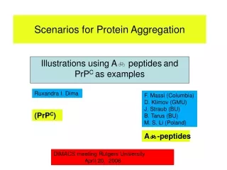
Ruxandra I. Dima F. Massi (Columbia) D. Klimov (GMU) J. Straub (BU) B. Tarus (BU) M. S. Li (Poland) Scenarios for Protein Aggregation Illustrations using A peptidesand PrPC as examples (PrPC) A-peptides DIMACS meeting Rutgers University April 20, 2006
Energy landscape for monomeric folding Monomer can misfold to multiple conformations Structural variations in the CBAs are imprinted in oligomers and fibrils
Aggregation Linked to diseases • Protein deposition diseases * transmissible spongiform encephalopathies (TSE; Mad Cow Disease) * Alzheimer’s disease, Parkinson’s disease * diabetes (type II) • All these diseases = related to misfolding and protein aggregation • Misfolding into multiple amyloid conformations (strains) • Examples: prion proteins (TSE), Alzheimer’s, CWD Question: What is the nature of the initial events in oligomer formation? Two broad scenarios: Illustrations using A peptides and PrPC Current AD hypothesis: Soluble oligomers are neurotoxic
Scenarios for Fibrillization (D.T., D. Klimov and R.Dima, Curr. Opin. Struct. Biol., 2003) A and TTR Prions N* = metastable N* formation = partial unfolding N* = stable N* formation in prions = unfolding of N KG depends on rate of formation of N* from N or U PrPc is metastable with respect to PrP* aggregation prone particle
Cascade of events to Fibrils Scenario I (Partial unfolding/ordering) Polydisperse Oligomers nA16-22 (A16-22)n
Differing Supra- molecular Assembly Heterogeneous Nucleation and Growth On + kM KG = F(Seq,C,GC) Heterogeneous Nuclei
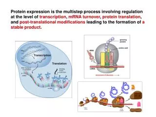
Ab-peptide in vivo is a metabolic product of precursor protein • Alzheimer’s Disease (AD) is responsible for 50% of cases of senile dementia • Ab-peptide is a normal byproduct of metabolism of Amyloid Precursor Protein (APP) • Cleavage of APP results from action of specific proteases called secretases Ab10-35 Ab1-40 and Ab1-42 peptides many naturally occurring mutants E22Q “Dutch” mutant • In Selkoe’s “Ab hypothesis,” AD is a result of the accumulation of Ab-peptide
A16-22 For Scenario I • Mechanism and Assembly Pathways • Sequence Effects • Role of water • Fragment has CHC • Interplay of hydrophobic/electrostatic effects
Trimer Structurefrom MD Antiparallel sheets Monomer is a Random Coil Structure: Inter-peptide Interaction Driven Interior is dry: Desolvation an early event
Dominant assembly pathway involves-helical intermediate Teplow JMB 2001 “Effective confinement” induces helix formation -helical intermediate “entropically” stabilized
Origin of -helical Intermediate Case I C C* Low Peptide Concentration C* = Overlap concentration Rjk ≈C-(1/3) Rjk Rjk/Rg 1 Polypeptide is mostly a random coil
C C* Peptides Interact Rjk / Rg ~ O(1) Peptide j is entropically confined j In peptide j confinement induces transient structure k For A16-22 interaction drives transient -helix formation
1 +NH3 COO- 2 -OOC NH3+ 3 +NH3 COO- Principle of Organization Anti-parallel registry satisfies Hydrophobic and charged interactions Hydrophobic andcharged residuesstabilize oligomers
Kinetics and stability of Oligomerization determined By balance of hydrophobic and Charged interactions Enhanced growth kinetics in E22Q due to change in charged states Massi,Klimov,DT, Straub (2002) Structural orientation requires charged residues “Long-range” correlations between charged residues in protein families linked to disease-related proteins (Dima and DT, Bioinformatics (2003) K16G/E22G trimer is unstable
Electrostatics interactions essential in amyloid formation: Charged states • E22Q “Dutch” mutant peptide shows enhanced rate of amyloid formation@ Ab10-35-NH2 Ab10-35-NH2E22Q • Lower propensity for amyloid formation in WT peptide due to Glu- charged states (versus Glno) • Proposed INVERSE correlation between charge and aggregation rate - now seen experimentally% *Zhang et al. Fold. Des. 3:413 (1998). @ Miravalle et. Al., J. Biol. Chem., 275, 27110-27116 (2000). #Massi and Straub, Biophys. J. 81:697 (2001); Massi, Klimov, Thirumalai and Straub, Prot. Sci. 11:1639 (2002). % Chiti, Stefani, Taddei, Ramponi and Dobson, Nature 424:805 (2003).
Tetramer forms rapidly Nucleus 4 Templated assembly Seed = Trimer Insert A16-22 monomer Barrier to addition
Important structural motifs in Ab-peptide monomer and fibrils • Ab-peptide structure determined in aqueous solution by NMR by Lee and coworkers* • Monomer Ab10-35peptide has well-defined “collapsed coil” structure • Collapsed coil is stabilized by VGSN turn region and LVFFA central hydrophobic cluster# central hydrophobic LVFFA cluster * S. Zhang et al., J.Struct. Biol.130, 130-141 (2000). # Massi, Peng, Lee and Straub, Biophys. J. 80:31 (2001). % Tycko and coworkers, PNAS 99: 16742 (2002). VGSN turn region
Scenario II (Global unfolding of PrPC) (D.T., D. Klimov and R.D., Curr. Op. Struct. Biol., 2003) A, TTR Prions N* = metastable N* formation = partial unfolding N = metastable N* formation involves global unfolding of N PrPSc growth kinetics Depends on rate of NN* transition KNN* KNN* depends on sequence and G† between N and N*
β* α Mechanism of assembly and propagation Prions • normal form PrPC = mostly a-helical • scrapie form PrPSc= mostly b-strand • the “protein-only hypothesis”: (Prusiner et al., Cell 1995 and Science 2004) PrPSc = template to catalyze conversion of normal form into the aggregate β Fluctuation Nucleation β β Growth PrPC* ? Propagation by recruitment
Question and Hypothesis Minimal infectious unit 90 121 231 Disordered in PrPC Ordered Proposal: PrPSc formation is preceded by transition from α PrPC* state Unfolded PrPC PrPC* ? PrPSc (48% β, 25% α) (45% α, 8% β) (20% α)
NMR Structure of Cellular form (PrPC) • Prions: “…Prion is a proteinaceous particle that lacks nucleic acid” (Prusiner, PNAS, 1998) • PrPC: 45% a, 8% b • PrPSc(90-231): 25% a, 48% b mPrPC(121-231) (Caughey et al. Biochemistry30, 7672 (1991)) Wuthrich 1997 (Cys179-Cys214)
H1 in mammalian PrPC is helical Charge patterns in H1 is rarely found in PDB, E. Coli and Yeast genomes
Random considerations: Pattern search for H1 in PrPC • (i,i+4) = oppositely charged residues • search sequences of 2103 PDB helices (Lhelix ≥ 6) (i,i+4) salt-bridges in mPrPC
Sequence analysis shows PrPC H1 is a helix • - X - - + X X + - X • search PDBselect (1210 proteins) • 23 (1.9%) sequences • 83% = α-helical, 17% = random coil • search E. Coli(4289 proteins) genome • 51 (1.2%) sequences • search yeast(8992 proteins) genome • 253 (2.8%) sequences Pattern of charged residues in H1 is unusual and NEVER associated with β-strand
Experiments and MD simulations show H1 is very stable Conformational fluctuations and stability of H1 with two force fields Stability is largely due to the three salt bridges in the 10 residue H1 from mPrPC
High helical propensity at all positions in H1 MOIL package (Amber and OPLS)(R. Elber et al.) H1 from mPrP (10 residues) positions 144-153 • 773 TIP3P water, 30 Ǻ cubic box, 300 K, neutral pH • 5 trajectories, 85 ns PDB Helix Strand
Unusual hydrophobicity pattern in H2 • X X X H H X X H H X H XH X X X X H P P P P X • search PDBAstral40(6000 proteins) • 12 (0.2%) sequences • the sequence is NEVER entirely α-helical (last 5 residues = non-helical in 87% of cases) • search E. Coli(4289 proteins) genome • 46 (1%) sequences • search yeast(8992 proteins) genome • 122 (1.4%) sequences Pattern of hydrophobicity of H2 is rare and NEVER entirely in a α-helix
H2+H3 in mammalian PrPC frustrated in helical state R. I. Dima and DT Biophys J. (2002); PNAS (2004) Conformational fluctuations in H2+H3 implicate a role for second half of H2 in the PrPC PrPC* transition
Structural transitions in H2+H3 NAMD package (Charmm) • H2+H3 in mPrP , S-S bond • H2 starts to unwind around position 187 • unwinding by stretching and bending
X-ray structure of PrPC dimer shows changes in H2 and H3 Domain-swapped dimer of huPrPC(Surewicz et al., NSB8, 770, 2001) • H1: 144-153 (monomer: 144-153) • H2: 172-188 and 194-197 (monomer: 173-194) • H3: 200-224 (monomer: 200-228) PDB file 1i4m
Rarely populated PrPC* shows changes in H2 and H3 15N-1H 2D NMR under variable pressure and NMR relaxation analysis on shPrP(90-231) (James et al., Biochemistry 41, 12277 (2002) and 43, 4439 (2004)) • in PrPC* C-terminal half of H2 and part of H3 are disordered 98.99% 1.0% 0.01%
Many pathogenic mutations are clustered around H2 and H3 From Collinge (2001) H2 and H3 region
Scenario for initiation of PrPC aggregation Finding: transition α PrPC* state initiated in second half of H2 and does not involve H1 G† Unfolded G† /KBT 1 PrPC PrPC* formation improbable PrPC* PrPSc (48% β, 25% α) (45% α, 8% β) (20% α)
Proposed structures for PrPC* PDB Amber and OPLS Charmm (48% α-helix) (30% α-helix) (20% α-helix) • H1 still α-helical • H3 only partially α-helical
Conclusions • Multiple routes and scenarios for fibril formation • Electrostatic and hydrophobic interactions determine structure and kinetics • Conformational heterogeneity in N* controls oligomer and fibril morphology (may be relevant for strains) • Phase diagram (T, C) plane for a single amyloidogenic protein is complex due to structural variations in the misfolded N* • Templated growth occurs by addition of one monomer at a time • Nucleus size and growth mechanism depends on protein