Download
1 / 20
210 likes | 514 Views
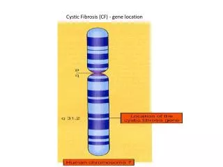
Cystic Fibrosis (CF) - gene location. Cystic Fibrosis (CF): Molecular defect. Cystic Fibrosis (CF): One gene may lead to many phenotypic effects. Cystic Fibrosis lungs. Lung from a CF patient, showing extensive destruction as a result of obstruction and infection. Normal lung.

E N D
Cystic Fibrosis (CF): One gene may lead to many phenotypic effects
Cystic Fibrosis lungs Lung from a CF patient, showing extensive destruction as a result of obstruction and infection Normal lung
Cystic Fibrosis Pancreas CF Pancreas showing infiltration of fat and fibrotic lesions Normal pancreas
Tay Sachs • Defective enzyme that breaks down a particular fatty substance
The hemoglobin tetramer BETA GLOBIN BETA GLOBIN Sickle cell mutation Sickle cell mutation Heme Heme Iron atom Iron atom ALPHA GLOBIN ALPHA GLOBIN
Sickle Cell Anemia (AR) Aggregation of hemoglobin molecules. Mutant hemoglobin molecules in red cells stack to form rodlike structures, which causes the red cells to deform.
Signaling cell Signaling molecule Plasma membrane 1 Receptor protein 2 Signal Transduction: How a cell can respond to signals from its environment Results in a change in which genes are expressed (turned on) 3 Target cell Fig. 11-12 Relay proteins Transcription factor (activated) 4 Nucleus DNA 5 Transcription mRNA New protein 6 Translation
Growth factor Receptor Target cell Hyperactive relay protein (product of rasoncogene) issues signals on its own Normal product of ras gene Relay proteins Fig. 11-20a Rasis an oncogene (cancer gene) the normal form of the gene is called a proto-oncogene Transcription factor (activated) DNA Nucleus Transcription Translation Protein that Stimulates cell division Oncogenes STIMULATE cell division
Proto-oncogene DNA Multiple copies of the gene Mutation within the gene Gene moved to new DNA locus, under new controls Fig. 11-18a New promoter Oncogene Hyperactive growth- stimulating protein in normal amount Normal growth- stimulating protein in excess Normal growth- stimulating protein in excess
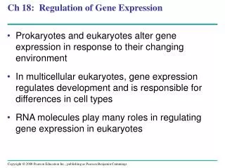
Normal tumor-suppressor genes prohibit cell division Growth-inhibiting factor Receptor Relay proteins Nonfunctional transcription factor (product of faulty p53 tumor-suppressor gene) cannot trigger transcription Fig. 11-20b Transcription factor (activated) Normal product of p53 gene Transcription Translation Protein absent (cell division not inhibited) Protein that inhibits cell division
Mutated tumor-suppressor gene Tumor-suppressor gene Fig. 11-18b Normal growth- inhibiting protein Defective, nonfunctioning protein Cell division not under control Cell division under control
A tissue comprised of billions of cells heterozygous for BRCA1 or BRCA2 Both alleles of BRCA1 or both alleles of BRCA2 must be mutant for cancer to develop. Why would in follow a dominant inheritance pattern? Your (my) probability of winning the lottery is very small. The probability that someone will win it is very large.