Download
1 / 42
530 likes | 894 Views
The Use of Synchrotron Radiation in Crystal Structure Analysis (Powder Diffraction). Al-Sharif Dept. of Physics Mu’tah University. Light matter interaction. *When light interacts with matter either, scattered (change direction) or absorbed .

E N D
The Use of Synchrotron Radiation in Crystal Structure Analysis(Powder Diffraction) Al-Sharif Dept. of Physics Mu’tah University
Light matter interaction *When light interacts with matter either, scattered(change direction) or absorbed. Both of these two interactions are useful to collect information about the matter. Scattering: Inelastic or elastic. *When light encounters an irregular material, scattering likely to be incoherent or random. *Ordered material (crystal) scattering produce a diffraction Pattern. Studying the resulted patterns reveals information about Matter.
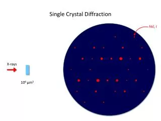
Light scattering and diffraction (a) Scattering by an atom (b) Diffraction by a crystal
Scattering from materials All materials scatter x-ray, even if they are not crystalline. Deviations from perfect periodicity spread the scattering out.
X-rays EM radiation with typical energies in the range 100 eV –100 keV. For diffraction applications, only short wavelengths x-rays (hard x-ray) in the range few angstroms – 0.1 angstrom are used. Because the wavelength of x-rays is comparable to the size of atoms, its suitable for probing the structure of a wide range of materials. Energetic x-ray can penetrate deep into the materials and provide information about the bulk structure.
WHY?? using the synchrotron radiationfor Diffraction Synchrotron facilities have become widely used as prefered source for x-rays diffraction High brightness: extremely intense (hundred of thousands of times than that of conventional x-ray tube). Highly collimated:small angular divergence of the beam. High energy: Short wavelengths high penetration. Highly polarized (linear or elliptical). Emitted in very short pulses: typically below a nanosecond). Low emittance.
Advantages for Synchrotron Diffractometer *Thehigh brightness and collimation of synchrotron beam, enable us to design an ultrahigh resolution detector on a perfect detector analyzer. *The broad band nature of the synchrotron radiation allows tuning the radiation through monochromators with special design to obtain an intense beam of several wavelengths .
Schematic diagram of powder diffraction system (NSLS,Brookhaven lab)
X-ray Diffractometer X-ray source sample Detector
Reflection and Transmission geometry of diffraction Solidsamples Liquidsamples
Powder Diffraction *Most widely non-destructive technique used(material characterization method ). *Powerful computers and 3rd generation x-ray synchrotron sourceshave transformed powder diffraction into a very powerful structural tool. *By measuring X-rays diffracted from the sample, one can obtain local structural information(phase identification ,strain, charge distribution, magnetic structure and texture,with an accuracy much higher than with standard diffraction.
Powder Diffraction • *Method is normally applied to data collected under ambient conditions. * Diffraction as a function of an external constraints (temperature, pressure, stress, electric field, etc.) can be done with specially designed sample containers.
Powder Diffraction *Various kinds of micro- and nano-crystalline materials can be characterized from x-ray powder diffraction including (inorganics, organics, drugs, minerals, zeolites, catalysts, metals and ceramics)
Powder Method *Crystal to be studied is reduced to a very fine powder and placed in a beam of monochromatic x-rays, (assemblage of smaller crystals oriented at random with respect to the incident beam). *The random orientation of powder equivalent to a single crystal rotated about all possible axes
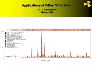
Bragg’s Law *In a randomly oriented crystalline powder, Bragg’s equation is simultaneously satisfied for large number of different (hkl) lattice planes. *The Powder Diffraction pattern of a sample represents a complete mapping of its structure.
Powder Diffraction *Qualitative Analysis Phase identification *Quantitative Analysis Lattice parameter determination Phase fraction analysis *Structure Solution and Refinement Rietveld method *Peak Shape Analysis Crystallite size distribution Microstrain analysis Defect concentration
Qualitative Analysis *The different phases of the studied sample can be identified from the Diff. Pattern. The diffraction pattern of known phases used to identify the phases present in the studied sample. *Computer Search match used to compare obtained pattern with ICDD data base of known compounds (over130,000 entries).
Quantitative analysis *We can determine the composition of the sample by measuring changes in the unit cell dimensions. *Rietveld method commonly used to determine the weigh fractions of multiphase mixtures.
Rietveld Refinement Goal: To obtain an accurate crystal structure. Basic Idea: To fit the entire diffraction pattern at once, optimizing the agreement between the calculated and observed patterns.
Powder diffraction *The observed diffraction line profile are distributions of intensities I(2θ) defined by several parameters, (i) Peak position (ii) Intensity (iii) width and shape
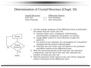
Steps to structure determination • Index the diffraction pattern. • Determine crystal system and unit cell dimensions. • Check for systematic absences to specify space group. • Do pattern fitting to get accurate unit cell dimensions and peak shape parameters. • Obtain a close structural model which allows atomic positions and displacement parameters to refine to optimize the fit to the observed diffraction (Rietveld refinement)
Information Obtained from Diffraction Pattern * Peak Positions Crystal system Unit cell dimensions Qualitative Phase identification * Peak Intensities Unit cell contents Quantitative Phase fraction Preferred orientation * Peak Shapes and Widths Crystallite size Strain Defects (dislocations, stacking faults, boundaries, etc.)
Crystallite size *As the crystallites in a powder get smaller the diffraction peaks in a powder pattern get wider. * Consider diffraction from a crystal of thickness t. How diffracted intensity varies as we move away from the exact Bragg’s angle????
Scherrer formula t=0.9λ/(B cosθ) Used to estimate the particle size of very small crystals(t) from the measured width(B)of their diffractioncurve θ: Diffraction angle
Extending Defects Extended defects disrupt the atomic arrangement of a crystal. These defect effectively terminate a crystallographically order domain of the crystal. Thus as far as x-rays are concerned one crystal ends and a new crystal begins at the extended defect. Crystallite size analysis on a sample containing extended defects can be used to estimate the ordered domain size (the size of the region between defects) Types of extended defects *Stacking faults (ABCABCABCCBACBACBA CBACBACBA) *Dislocations in layered materials (graphite, MoS2, clays,……..) *Antiphase boundaries, which arise in partially ordered boundaries materials (Cu materials (Cu3 Au, Sr2 AlTaO6))
Peak Broadening Other sources affect peak broadening (beside crystallite size) must be taken into consideration when analyzing diffraction data: *X-ray beam is not parallel. • * X-ray beam is not perfectly Monochromatic * Strain.
Strain Strains may exist in material. It vary from grain to grain or within a grain (microstrain - nonuniform) leads to systematic shifts of atoms from the ideal positions. Theremayalso be auniform strain due to an external load (macrostrain), causes the unit cell to expand or contract in an isotropic way, leading to changes in the unit cell parameters influence the diffraction pattern. * Microstrainsproduces peak broadening. * Macrostrainproduces peak shift.
Effect of strain in diffraction Peaks Macrostrain produces peak shift. Microstrains produces peak broadening.
Microstrain broadinning Micro-strain leads to a distribution of d-spacing in crystal. This can be evaluated by differentiating Bragg’s law -B = Δ2θ = -2 (Δd/d) tanθ B is the extra broadening over and above that present due to the instrument resolution and particle size. We can calculate microstress from microstrain using elastic modulus.
Diffraction intensity The positions of the atoms in the unit cell affect the intensities but not the directions of the diffracted beam.Atomic positions can be determined by observations of intensities.
Structure factor (F) amplitude of wave scattered by all atoms of a unit cell F = ______________________________________________ amplitude of wave scattered by one electron F (hkl) = Summation extend over all the N atoms of the unit cell f: atomic scattering factor u, v, w:are the fractional coordinates
Lorentz – polarization factor The overall effect of these factors is to decrease the intensity of diffraction at intermediate angles.
Temperature factor *Atoms are not fixed at lattice points. Atoms undergo thermal vibrations about their main position. The amplitude of its vibration increases with temperature (unit cell expand). *Thermal agitation smear out the lattice planes decreasing the intensity. *Thermal vibration of atoms causes some general coherent scattering In all directions increasing the background gradually with θ.
Recommendation These are some of the many things that we can extract from powder diffraction technique concerning the crystal structure analysis. I suggest a one to two days workshop must be planned in the near future that should be devoted fully to every technique that its planned to be used in the synchrotron facility.