Download
1 / 11
260 likes | 882 Views
Nuclear Medicine Project Physics 10. Mayur Bansode Evelyn Cuevas Stephanie Merlos Paul Mitchell Brian Wolter Mike Zamora 15 November 2010. The History. Herman Blumgart 1925- First use of radioactive tracers for diagnosis. Hal Anger

E N D
Nuclear Medicine ProjectPhysics 10 Mayur Bansode Evelyn Cuevas Stephanie Merlos Paul Mitchell Brian Wolter Mike Zamora 15 November 2010
The History Herman Blumgart 1925- First use of radioactive tracers for diagnosis. Hal Anger 1958- presented his first scintillation camera which led to cardiology. Henri Becquerel 1896-Discovered mysterious “rays”. 1903-Nobel Prize Marie Curie 1897- named mysterious rays “radioactivity” 1903-Nobel Prize
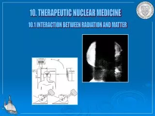
How It Works Viewing Internally w/out invasiveness? Radioactivity- Alpha, Beta Particles. Gamma Rays. Radionuclides- unstable nucleus. Injected, Ingested, Inhaled. Once in body, localizes specific organs or cell types.
99mTc, Gallium 67, and 210Tl PET, CT, SPECT scans Therapeutic Procedures New techniques, Combining.
What is a PET Scan? Medical diagnostic procedure which produces 3D or 4D images in a region of the body. How Do PET scans work? PET scans distribute small amounts of radioactive material into a desired region of the body by intravenous injection. The specified region of the body is than scanned and an image is made by showing where or where not the material is being metabolized. How is the image produced on a screen? The scanning of the body region which was injected with the radioactive material produces millions of lines where the Positrons are detected in the body. The lines are than put together on a screen to create a 3D image. Positron Emission Tomography
What are some common applications of the PET scan? • Oncology • PET scans are commonly used to produce images of tumors in the body, and help diagnose types of cancers. • This is the most common use of the PET scan • Neurology • PET scans make it helpful to treat and diagnose neurological disorders such as stroke, epilepsy, Alzheimer’s and brain tumors. • Cardiology • PET scans can measure the amount of blood flow to an area of the heart. Making it easier to treat and diagnose heart disease. • What is the future of PET scans? • PET scans are becoming more powerful and more precise. • Physicians are discovering new ways of treating and diagnosing medical ailments.
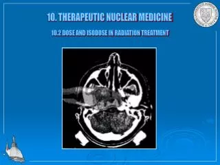
Computed Tomography ( CT Scan) • The primary goal of any CT system is to accurately reproduce the internal structures of the body as two-dimensional cross-sectional images. • This goal is accomplished by computed tomography's superior ability to overcome addition of structures and demonstrate slight differences in tissue contrast. • It is important to realize that collecting many projections of an object and heavy filtration of the x-ray beam play important roles in CT image formation. • Each component of a CT system plays a major role in the accurate formation of each CT image it produces.
A CT Scan an upgraded version of an X-ray combined with computer technology. The result is a clearer, more-detailed view of the inside the body. CT scans can produce pictures of an individual's bones, organs like the kidneys and even blood vessels. Common Uses Chest (Thorax): Look for problems in the lungs Abdomen: To diagnose cysts, tumors or infections For Urinary Tract infections: Kidney Scan for stones Liver: To find Liver tumors or bleeding Pancreas: Inflammation of the pancreas Gallbladder: Stones Pelvis: To look for problems in the pelvis like ovarian cysts Arms and Legs: To find problems in extremities
Future in Nuclear Medicine • While hybrid or multimodality imaging continues to evolve and increase in popularity, spurred by evidence-based medicine, by 2020, it won’t matter if it is PET/CT, SPECT/CT, or PET/MR or even SPECT/MR—the border between the imaging modalities will have disappeared. • Instead of a modality-specific focus, nuclear medicine in the future will be integrated into organ-specific or disease-specific groups. • Patients will be reaping the rewards of the transition away from a modality-specific focus to more image fusion within nuclear medicine. “The future will be more and more image fusion so that physicians won’t think so much about it being PET, CT or MRI. Instead, for each individual patient, it will be a more tailored, more individualized protocol using functional imaging with anatomical imaging, regardless of which modality you choose. • Utilization of in vivo and in vitro markers will play a vital role in nuclear medicine. • In vitro tests—which include the genetic analysis of blood samples—will be much more important to identify high-risk patient populations, such as individuals at risk for developing cardiovascular disease or cancer. • The future also could bring a scanner that can do more than two modalities at the same time. For example, a patient comes in for a blood test; three imaging tests are performed along with a non-invasive biopsy which will provide better tumor characteristics than we currently obtain now. • By combining the individual genetic and chemical workup of each patient, with imaging and data, such as age, sex, weight, disease susceptibility and family history, “we will see a whole explosion of how patients are diagnosed in the future.
Summary • Nuclear medicine is a branch of medicine that uses the nuclear properties of matter in diagnosis and therapy. More specifically, it is a part of molecular imaging because it produces images that reflect biological processes that take place at the cellular and subcellular level. • Additionally, nuclear medicine imaging like PET Scans or CT Scans is a dataset of one or more images that has the ability to slice through the patient at a particular position with a rotating gamma-camera. • Lastly as nuclear medicine continues to make huge advances in dataset imaging and genetic tracing, doctors will have the ability to identify and treat patient quicker and possibly eradicate some diseases all together. As a result, healthcare cost may stabilize or even decrease making healthcare affordable for everyone.
References Dr. Brian Renner from Global Holdings Imaging “Henri Becquerel-Biography”. Nobelprize.org. n.d. web 13 Nov 2010 http://nobelprize.org/nobel_prizes/physics/laureates/1903/becquerel-bio.html “Important Moments in the History of Nuclear Medicine”. SNM. n.d. web 13 Nov 2010 http://interactive.snm.org/index.cfm?Page.ID=1107 Nuclear medicine Information. SNM n.d. web 13 Nov 2010 http://www.bookrags.com/wiki/Nuclear_medicine Wagner, Henry N., Jr. MD. “Nuclear Medicine: 100 Years in the Making”. The Journal of Nuclear Medicine.Vol.37. 10 October 1996. web 13 Nov 2010 http://jnm.snmjouranls.org/cgi/reprint/37/10/18N.pdf. http://www.webmd.com/a-to-z-guides/computed- tomography-ct-scan-of-the-body www.cancerresearch.com www.righthealth.com www.petscaninfo.com