Download
1 / 31
310 likes | 334 Views
This article explores genome-wide analysis using molecular karyotyping techniques such as oligo/SNP arrays. It discusses chromosomal abnormalities, their prevalence, indications for chromosome study, and various methods for analysis. The advantages and disadvantages of karyotyping, FISH analysis, and array-based comparative genomic hybridization are also presented. Furthermore, the article highlights the different array formats and their uses, including high-resolution BAC-based arrays, oligo-based arrays, and SNP arrays. Three case studies illustrate the application of molecular karyotyping in diagnosing developmental delays, intellectual disabilities, and other genetic disorders.

E N D
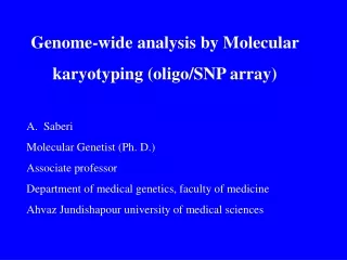
Genome-wide analysis by Molecular karyotyping (oligo/SNP array) A. Saberi Molecular Genetist (Ph. D.) Associate professor Department of medical genetics, faculty of medicine Ahvaz Jundishapour university of medical sciences
Chromosomal Abnormalities • Numerical • Extra or missing chromosomes (aneuploidy) • Polyploidy • Mosaicism • Structural • deletions/duplications, • translocations, • inversions, etc.) Most cancers acquire structural or numerical chromosomal abnormalities
Chromosomal Abnormalities • Are common • 1% of live birth • 2% of pregnancies in women > 35 • 50% of all spontaneous 1st trimester abortions
Indications for chromosome study • Problems of early growth and development: failure to thrive, developmental delay, dysmorphic faces, multiple malformations, short stature, ambiguous genitalia and MR. • Stillbirth and neonatal death • Fertility problems: couples with a history of infertility or multiple pregnancy loss, women with amenorrhea • Family history: a known/suspected chr. abnormality in a first degree relative • Neoplasia • Pregnancy in a woman of advanced age (>35 yrs)
Chromosome analysis methods: 1. Karyotype
High resolution karyotype Advantages “Whole genome scan” Relative low cost Disadvantages Labor intensive Detection above 5 Mb
2. FISH probe for chromosme 15
FISH Analysis Advantages Highl resolution Used for both metaphase and interphase cells Disadvantages Time consuming Is not genome-wide screening Can not detect unexpected imbalances 500-600 probes needed to match the power of karyotyping
To improve detection… Additional molecular cytogenetic technologies are needed that offer a whole genome approach to submicroscopic imbalances
Array-based Comparative Genomic Hybridization Molecular cytogenetic method to detect copy number imbalances Higher resolution and throughput Possibilty for automation, robustness and simplicity Precise mapping of aberrations No need for long-term cell culture and less labor intensive Whole genome scaning Reveals clinically unsuspected genomic imbalances Detect genomic duplications that cannot be identified by FISH Objective method compared to routine cytogenetic G-banding and FISH analysis
Indications Global developmental delay Mental retardation Autism Multiple congenital anomalies Dysmorphism Powerful tool in disease gene discovery Powerful tool in prenatal diagnosis Promising for cancer research (diagnosis, classification and prognosis)
Array FormatsHigh Resolution BAC based array –detection of CNVs with low resolution Oligo based array-detection of CNVs with higher resolution SNP array- detection of CNVs + genotyping, UPD and LOH
Normal Deletion Duplication A A A A A A B B B A A A B B B B B B A A A B B B A B B B A A A B AAA AA AA A AAB AB AB ABB B BB BB BBB Deletion Normal Duplication Normal
SNP arrays more sensitive for detection of mosaicism Non-mosaic deletion Mosaic deletion
Long continuous stretches of homozygosity (LCSH) with normal copy number Small deletion SNPs and consanguinity or UPD Chromosome 2
case #1 • A 10 years girl Developmental delay, • Parents: second cousin growth retardation • Normal pregnancy, delay in speech • Birth weight: 3.25kg Begins to walk at 3 years • hypotonia, Intellectual disability • Seizure is controlled with medication • Brain MRI: normal
Main features of 3q13.31 deletion syndrome • Low muscle tone (hypotonia), so a baby feels floppy to hold • Developmental delay, including mobility and speech • Variable need for support with learning • Above average growth rate in babies and children, including head size • Small genitals in boys. Genitals in girls are normal • Characteristic facial features • High palate (roof of the mouth) • Less common features • Brain and central nervous system involvement • Seizures and/ or unusual patterns of electrical activity in the brain • Eyesight problems including short or long sight • (Molin 2012; Shuvarikov 2013)
Case #2 A 6 years boy, developmental delay, speech problem (beginning at 2.5 years, after 6 years very simple sentences), narrow face, flat mid face, long neck, seizure, MRI (probability of demyelination in subcortical frontoparietal lobes), iron deficiency, mental disability, attention deficit disoreder, normal health parents, noconsanguineus parent, normal metabolic test normal fragile-X test
2.8 Mb, 6p23 -6p22.3, 11 RefSeq. Genes , ATXN1 Our patient 5.4 Mb 6p22.3-p23, 21 RefSeq genes, ATXN1 Log2 ratio 1 Mb 6p22.3, ATXN1 14.6 Mb, 6p22.3-p24.3 58 RefSeq genes, ATXN1 3.6 Mb, 6p23-p24.3 , 28 RefSeq genes, ATXN1 5.2 Mb, 6p22.3 18 ,RefSeq genes, ATXN1 8.8 Mb, 6p22.3-p24.1, 34 RefSeq genes, ATXN1 chromosome 6 position
Case #3 • A 5years boy • Non-caonsanginuous parent • Cesarean delivery • Birth weight: 2.7kg • Growth retardation • Developmental delay • Begin to speak at 3 years, • nasal speech • Seizure during fever • Intellectual disability
Case #4 • AGE : 4 YEARS • Birth weight: 1.700g • Developmental delay: delay in speech (only few words at this age), delay in walking, • Seizure, VSD, hypothyroidism, malform teeth • Normal MRI • Normal karyotype • No sisters or brothers • Unconsanguinous parents
Del 5.6Mb 12p13.33-12p13.31 , 32 OMIM genes, Timoty Brugada syndrome Dup 17.6Mb 16q22.3-16q24.3 80OMIM genes, DD and MR