Download
1 / 21
290 likes | 1.09k Views
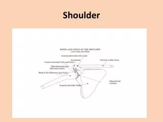
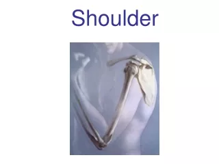
Shoulder Ultrasound. Personal protocols Caitlin Gardiner. Introduction.

E N D
Shoulder Ultrasound Personal protocols Caitlin Gardiner
Introduction • The practice I did the majority of my training with in my first year of clinical ultrasound performs little MSK ultrasound. I have just commenced work in the past few weeks at a general radiology practice where I am already exposed to a significant degree more MSK ultrasound that ever before. At this stage, I’m scanning MSK with another sonographer and the plan is to slowly add various scans to my list as I gain competency. Shoulder ultrasound is challenging due to the large number of tendons very proximal to one another and unique scanning plane. Using my images I submitted for homework, this is a summary of what I know so far with the feedback I have been given by my tutor.
Biceps Patient Position/Manoeuvre Assessment of Structure • Ipsilateral hand of the shoulder placed on the knee with palm upwards. • Observe in Trans • -?surrounding fluid • Observe it Long • ?regular fibers • Any hypoechoic/thickening • Ensure visualization from upper biceps muscle to very superior segment • Assess groove • <3mm= shallow • Transverse bicep central/medial edge(subluxed)/out of the groove (dislocated) • Assess pop-eye sign, to aid biceps rupture
Subscapularis Patient Position/Manoeuvre Assessment of Structure • Place probe in transverse, fully rotate the arm externally • Assess subscupularislike this and then visualize in mild extension and partial external rotation • Assess all fibres, including those adjacent to the bicep tendon (utilise manoeuvres) • Ensure observation of subscapularis sliding under coracoid (?entrapment) • Visualise fibrillar pattern and any bursal thickening • Mild bony irregularly is acceptable
Submitted Images and Feedback Image with internal rotation does not have subscapularis in it
CA Ligament Patient Position/Manoeuvre Assessment of Structure • Maintain external rotation of the arm. Slide the probe medial to visualize the clavicle and angle the lateral end of probe superior to around 45° to see acromium.
AC Joint Patient Position/Manoeuvre Assessment of Structure • ‘Plonk’ probe on top, in line with the clavicle • Is there any focal tenderness? • Any separation of the two bones (can get patient to pull from underneath of bed to apply pressure) • Any cysts or bony spurs
Submitted Images and Feedback Lovely.
Supraspinatus Patient Position/Manoeuvre Assessment of Structure • Place the ipsilateral hand on the ipsilateral hip with elbow posterior. Visualize the bicep in transverse in the most medial side of the screen. Slide the probe backwards to visual is transverse. Rotate 90°to view tendon in longitudinal. • Consider various positions (eg, hand behind back/neutral) throughout to maximize assessment • In true transverse, assess from anterior to posterior ensuring visualisation adjacent to biceps tendon • Regular contour (?thinning, flattening) • In longitudinal, image from medial to lateral • fibrillar pattern • Bony irregularity (?enthesopathy) • Bursal thickening • Note any calcifications and associated hypervascularity
Submitted Images and Feedback None of trans images have biceps in them, therefore most anterior portion is not imaged. Mid and post long images are a little oblique.
Infraspinatus Patient Position/Manoeuvre Assessment of Structure • Place hand on contralateral shoulder. Position probe on posterior of shoulder (not too far back). • Mild bony irregularity is acceptable • Significant bony irregularity/Hill-Sachs deformities indicate dislocation • If need to differentiate from supraspinatus, refer to previous position where the infraspinatusfibres run oblique
Submitted Images and Feedback Lovely.
Posterior Joint Patient Position/Manoeuvre Assessment of Structure • Drop field of depth from infraspinatus and slide probe slightly medial • Ask patient to slowly tap ipsilateral shoulder and observe any joint fluid. • Appears as an echogenic triangle • ?fluid • ?cysts
Submitted Images and Feedback No feedback.
Spinoglenoid/Suprascapular Notches Patient Position/Manoeuvre Assessment of Structure • Spinoglenoid notch- move probe more medially • Suprascapular notch- position probe between the superior scapula and the posterior border of the lateral clavicle • Exclude ganglion and cysts
Submitted Images and Feedback ?Mislabelled
Abduction Patient Position/Manoeuvre Assessment of Structure • Patients arm is bent beside their torso with their palm up. Abduct slowly. • Observe anterior and mid supraspinatus under CA Ligament • ? Bunching of the supraspinatus • ?bunching of the bursa
References • Ideas extracted from • McNally E, 2005. Practical Musculoskeletal Sonography. Elevisier Churchill Livingstone, Philadelphia • Coombs P, 2005. Shoulder Ultrasound: a discussion paper. Soundeffects; 3:18-25