Download
1 / 80
890 likes | 1.35k Views
Hypothermia for Hypoxic Ischemic Encephalopathy. Mitchell Imm, M.D. What now?. Meta-analysis (3 randomized clinical trials) shows that mild hypothermia is associated with a significant reduction in death or severe disability

E N D
Hypothermia for Hypoxic Ischemic Encephalopathy Mitchell Imm, M.D.
What now? • Meta-analysis (3 randomized clinical trials) shows that mild hypothermia is associated with a significant reduction in death or severe disability Edwards and Azzopardi. “Therapeutic Hypothermia Following Perinatal Asphyxia.” Archives of Disease in Childhood: Fetal and Neonatal ed. 2006. • What form: Selective head cooling vs. Whole Body cooling? • What degree of hypothermia provide the most neuroprotection? • What is the optimum duration of hypothermia? • Is it effective outside of 6 hours? • Is it protective for infants with severe HIE? • Will the benefits endure when the child reaches school age?
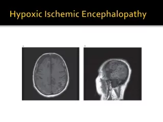
Hypoxic Ischemic Encephalopathy • Causedby impaired cerebral blood flow • Moderate to severe HIE occurs 2-4/1000 live births • 10% mortality rate for newborns with moderate HIE • 60% mortality rate for newborns with severe HIE. • 25-30% survivors with moderate HIE have long term disabilities • Nearly 90% survivors with severe HIE have long term disabilities
Hypoxic Ischemic Insult • Usually cause by interruption in placental blood flow • Subsequent interruption of cerebral blood flow • fetal response • Cerebral vasodilatation • Redistribution of organ blood flow • Loss of cerebral autoregulation
Birth Asphyxia • Condition of impaired gas exchange that leads to hypoxemia and hypercapnia • Fetal acidosis: umbilical arterial pH <7.0 and base deficit >=12 • Early onset of moderate-severe encephalopathy • Spastic quadriplegic or dyskinetic CP • Exclusion of other identifiable etiologies • Other nonspecific criteria • Sentinel hypoxic event before or during labor • Sustained fetal bradycardia or other signs of non-reassuring fetal status • APGAR 0-3 beyond 5 minutes • Multi-organ failure within 72 hours • Early imaging studies showing acute, non-focal cerebral abnormality • From ACOG Task Force in Neonatal Encephalopathy and Cerebral Palsy, 2003
Risk Factors • Severe preecclampsia • Placental abruption • Multiples • Antepartum hemorrhage • IUGR • Malpresentation • Cord prolapse • Stat C-section • Maternal fever
Primary Energy Failure • Decreased high energy phosphate compounds, e.g. phosphocreatine and ATP • Failure of ion pumps leading to Na, K, Ca ionic gradient disturbances • Mitochondrial dysfunction • Glutamate release • NMDA receptor overstimulation
Reperfusion • Return of cerebral blood flow • Normal blood pressure • Normal intracellular pH • Transient improvement of cytotoxic edema • No seizures
Latent Phase • Depressed EEG activity • Recovery of the mitochondria
Secondary Energy Failure • 6-24 hours after insult • Decreased high energy phosphates • Similar pathways as Primary Energy failure • Seizures • Cytotoxic edema • Excitotoxins • Cell death
Secondary Energy Failure “The severity of secondary phase energy failure is strongly correlated with adverse neurodevelopmental outcomes at 1 and 4 years of age.” RA Polin, TM Randis, R Sahni. “Systemic Hypothermia to Decrease Morbidity of Hypoxic-ischemic Brain Injury.” Journal of Perinatology. 2007
Mechanisms of Cell Injury • Failure of ion pumps • Influx of Na, Ca, and water • Glutamate release and subsequent activation of NMDA glutamate receptors • Further increase in intracellular Ca • Activation of lipases, proteases, endonulceases, phospholipases • NO synthesis • Oxygen free radical synthesis • Free fatty acid peroxidation • Inflammatory mediators
Cell Death • Necrosis • organelle disruption • plasma membrane disruption and rupture • cell swelling • Apoptosis • programmed cell death • cell shrinkage • nuclear pyknosis • chromatin condensation • genomic fragmentation
Apoptosis • Caspase activation • cysteine proteases • cleavage of inhibitor of caspase-activated DNase • Inactivates DNA repair enzyme • activation of caspase-activated DNase • DNA fragmentation and chromatin condensation • Apoptosis inducible factor • independent of caspase • translocates to the nucleus • leads to DNA fragmentation and chromatin condensation
Patterns of Brain Injury • Most common area of injury occurs in parasagittal cortex (watershed areas) and basal ganglia and thalamus. • Parasagittal injury caused by prolonged or partial asphyxia and causes cognitive impairments • Basal ganglia/thalamus injury caused by acute, near total asphyxia and presents with more seizure activity. Long term sequelae are cognitive defects, rigidity, seizures, motor speech impairment
Modified Sarnat Staging of HIE • Level of consciousness • Activity • Neuromuscular control • Complex/primitive reflexes • Autonomic function • Seizures
Sarnat Stage 1 (mild) • Hyper-alert • Active • Normal muscle tone • Mild distal flexion • Overactive stretch reflexes • Weak suck • Strong Moro • Slight tonic neck • Mydriasis • Tachycardia • No seizures • Neuro exam usually normalizes by 3-4 days • No long term sequelae
Sarnat Stage 2 (moderate) • Lethargic or obtunded • Decreased activity • Mild hypotonia • Strong distal flexion • Overactive stretch reflexes • Weak or absent suck • Weak or incomplete Moro • Strong tonic neck • Miosis • Bradycardia • Focal or multi-focal seizures
Sarnat Stage 3 (severe) • Stupor or coma • No activity • Flaccid muscle tone • Intermittent decerebration • Decreased or absent stretch reflexes • Absent suck • Absent Moro • Absent tonic neck • Variable pupils; fixed, deviated, non-reactive, and dilated • Variable heart rate • Uncommon seizures
Other Studied Treatments • Mannitol • Glucocoticoids • Phenobarbital • Calcium Channel Blockers • Magnesium Sulfate • Allopurinol • Resuscitating with FiO2 0.21 instead of 1.0 • Superoxide dismutase
Hypothermia for HIE • Miller et al. “Hypothermia in the Treatment of Asphyxia Neonatorum.” 1964 • Gluckman et al. “Selective Head Cooling with Mild Systemic Hypothermia after Neonatal Encephalopathy Multicenter Randomized Trial.” Lancet. 2005 • Shankaran et al. “Whole Body Hypothermia for Neonates with Hypoxic-Ischemic Encephalopathy.” New England Journal of Medicine. 2005.
Hypothermia for HIE • in animal models temperature reductions of 2-5 degrees C provided neuroprotection • Impacts multiple pathways to brain injury • Affecting excitatory amino acids, brain metabolism, cerebral blood flow, nitric oxide production, apoptosis
Selective Head Cooling • N=234 randomized. 218 subjects were evaluated at 18 months • 07/1999 to 01/2002 • Cool cap placed within 6 hours (avg. 4.3 hours of life) • Used the Cool Cap from Natus Medical Incorporated. http://www.natus.com • Infants randomized to normothermic control group (rectal temp 37 degrees Celsius) or hypothermic treatment group (rectal temp 34 degrees Celsius) for 72 hours
Selective Head Cooling • Follow up @ 18 months for a neurodevelopmental exam; height, weight, head circumference measurements; Bayley-II psychometric testing; audiology assessment; vision assessment • Primary Outcome measures: Mortality and Severe Neurodevelopmental Disability (Gross Motor Function impairment level 3-5, Bayley mental scale<70, bilateral cortical visual impairment)
Selective Head Cooling • entry criteria • At least 36 weeks gestation • APGAR <5 @ 10 min, or • Continued resuscitation, or • Severe acidosis ph<7, base deficit >= 16 on an umbilical cord blood sample or ABG/VBG done within 60 minutes of birth • Then, assessed for moderate or severe encephalopathy using the modified Sarnat • Then, aEEG showing moderate or severe background activity and/or seizures
Normal aEEG Upper margin of band of aEEG activity above 10 uV and lower margin of band of aEEG activity above 5 uV
Moderately abnormal aEEG Upper margin of band of aEEG activity above 10 uV and lower margin below 5 uV.
Severely Abnormal aEEG Upper and lower margin of band of aEEG activity below 10 uV
Seizures on aEEG sudden increase in voltage accompanied by narrowing of the band of aEEG activity and followed by a brief period of suppression
Selective Head Cooling • Adverse Events • Overall there was no statistically significant difference between the treatment and control groups, except for: • Minor cardiac arrhythmia • 9% vs. 1% • Mild sinus bradycardia • Scalp edema • 21% of treatment subjects • Resolved without any intervention after Cool Cap removed
Treatment group 108 subjects 45% (49) favorable outcome 55% (59) unfavorable outcome Death 33% Severe motor disability 19% Control group 110 subjects 34% (37) favorable outcome 66% (73) unfavorable outcome Death 38% Severe motor disability 31% Results No significant difference in rates of death or severe disability at 18 months (p=0.10)
Results • Infants with moderate aEEG abnormalities (N=172) there was a significant reduction in death or severe disability @ 18 months (48% vs. 66%, p=0.02) • Infants with severe aEEG abnormalities (N=46) there was no significant difference (68% vs. 79%, p=0.5)
Conclusions “Although induced head cooling is not protective in a mixed population of infants with neonatal encephalopathy, it could safely improve survival without severe neurodevelopmental disability in infants with less severe aEEG changes.”
Whole Body Hypothermia • N=208 (239 eligible) • 07/2000 to 05/2003 • Patients enrolled by 6 hours of life (avg. 5 hours 2 min of life) • Subject randomized to hypothermic treatment group (esophageal temp 33.5 degrees C) for 72 hours vs. normothermic control group
Methods • Treatment group placed on infant blanket cooled to 5 degrees C. Esophageal temperatures were monitored and set to 33.5 degrees C by the servomechanism. Abdominal wall temperatures monitored. • Esophageal and abdominal wall temp monitored every 15 min for 4 hours, every hour for 8 hours, then every 4 hours • Rewarming 0.5 degrees C every hour till infant’s temp was 36.5 degrees C after 72 hours of hypothermia
Follow up • Primary outcome: death or disability (moderate or severe) • Evaluated at 18-22 months for neuromotor disability, Bayley II, growth, vision assessment, hearing assessment • Severe disability defined as Bayley Mental Development Index<70, GMFCS grade 3-5, hearing impairment requiring hearing aids, blindness • Moderate disability Bayley MDI 70-84 plus 1 of the following: GMFCS grade 2, epilepsy, hearing impairment without amplification
Entry Criteria • At least 36 weeks gestation • Acidosis pH<7, base deficit >=16 on an umbilical cord blood sample or 1 hour blood gas, or • If pH 7.01-7.15, base deficit 10-15.9, and blood gas unavailable, there must be acute perinatal event and 10 min APGAR <= 5 or assisted ventilation for >= 10 min • Then, neurological examination to determine severity of encephalopathy using the modified Sarnat
Sarnat Stage 2 (moderate) • Lethargic or obtunded • Decreased activity • Mild hypotonia • Strong distal flexion • Overactive stretch reflexes • Weak or absent suck • Weak or incomplete Moro • Strong tonic neck • Miosis • Bradycardia • Focal or multi-focal seizures
Sarnat Stage 3 (severe) • Stupor or coma • No activity • Flaccid muscle tone • Intermittent decerebration • Decreased or absent stretch reflexes • Absent suck • Absent Moro • Absent tonic neck • Variable pupils; fixed, deviated, non-reactive, and dilated • Variable heart rate • Uncommon seizures
Exclusion Criteria • Unable to enroll by 6 hours of age • Major congenital anomalies • Severe growth restriction (BW<1800 grams) • Refusal of consent • Moribund infants for whom no further treatment was planned
Adverse Events • no significant difference between treatment and control groups, except for sinus bradycardia and skin changes • Mean HR 109 for treatment group vs. 140 for control group • Skin changes include erythema, sclerema, cyanosis, subcutaneous fat necrosis (4/102 infants)
Treatment group 102 subjects 56% (57) favorable outcome 44% (45) unfavorable outcome Death 24% (24) Control group 103 subjects 38% (39) favorable outcome 62% (64) unfavorable outcome Death 37% (38) Results Significant difference in rates of death or moderate to severe disability at 18-22 months (p=0.01).
Results • There was no significant difference between treatment and control groups when basing it on the degree of encephalopathy (moderate 32% vs. 48%, p=0.09; severe 72% vs. 85%, p=0.24) • There was no significant difference when comparing the incidence of disabling CP, blindness, severe hearing impairment, Bayley Mental Development Index, Bayley Psychomotor Development Index