Download
1 / 53
550 likes | 818 Views
Teledermatology and Telepsychiatry. Dr M N Kamel Boulos MIM Centre, City University, London E-mail: M.Nabih-Kamel-Boulos@city.ac.uk. Teledermatology and Telepsychiatry. Part 1: Teledermatology and Telepsychiatry in a Nutshell

E N D
Teledermatology and Telepsychiatry Dr M N Kamel Boulos MIM Centre, City University, London E-mail:M.Nabih-Kamel-Boulos@city.ac.uk
Teledermatology and Telepsychiatry Part 1: Teledermatology and Telepsychiatry in a Nutshell Part 2: A Practical Guide to Teledermatology Imaging and Evaluation
Teledermatology and Telepsychiatry Part 1 Teledermatology and Telepsychiatry in a Nutshell
Teledermatology: Introduction • Dermatology involves various activities such as talking with the patient to elicit a history, visual pattern recognition, surgical treatment and pre- and postoperative management. • Teledermatology, the use of telemedicine for dermatological consultations, can serve as a good model for other telemedicine specialities. In particular, a key element in making a dermatological diagnosis, visual inspection of the skin (dermatology is a very visual medical speciality), can be readily transmitted via telemedicine techniques.
Teledermatology: Benefits • The majority of dermatologists practice in or around urban areas, which leaves rural areas with fewer specialists. Teledermatology can be a means of delivering specialist care to these out of reach patients. This is very important because of the fact that many general practitioners are unable to diagnose even the most common dermatological diseases. • During a teledermatological consultation, a dermatologist will evaluate clinical and laboratory data, make a diagnosis, and prescribe therapy for patients located at a distance. Ultimately, the goal is to provide care to dermatology patients in underserved areas, improve the quality of care, and decrease the cost of care.
Non-Dermatologists Treatment of Skin Diseases • Studies have shown that non-dermatologists perform poorly in the diagnosis and treatment of skin disease. • Low concordance in diagnosis (41% for medical attending physicians) with that of dermatologists and a high number of inappropriate skin biopsies. J Am Acad Dermatol (32), 5, pt.1, 726-729; 1995 • 400 dermatologist and non-dermatologists asked to identify 20 common dermatological disorders from colour transparencies. • Internal medicine residents - 49% correct • Internal medicine staff - 52% correct. Arch Dermatol (117): 620-622; 1981
Teledermatology: Problems • Although promising, teledermatology does also have its problems. The first and major disadvantage is the inability of the consulting physician to physically examine the patient’s skin (physical examinations are a major way the dermatologists evaluate and treat patients). For example, in one case, the consulting physician is unable to palpate the skin, and as a result, the doctor is unable to make a fully informed decision. • Secondly, the lack of proper reimbursement schemes in some countries inhibits the growth of teledermatology.
Teledermatology: Types - 1 • Teledermatology programs can be deployed using either live-interactive video (real-time or synchronous transmission) or store-and-forward technology (pre-recorded or asynchronous). • The major advantage of the first technique (live interactive technology) is that the consulting physician can ask the patient and referring physician any questions that may be essential to making a diagnosis and carrying out treatment.
Teledermatology: Types - 2 • The store and forward technology is more convenient due to the fact that there is no need of a consulting physician at the same time as the referring physician and patient encounter. Compared to the other type, the store and forward method is also time efficient. • However, this method does have its disadvantages. The consulting physician can not ask the patient any questions and he cannot ask for certain images or close-up images. This can be very problematic because it prevents the consulting physician from making a better diagnosis. • Both technologies use two very useful instruments, a dermatoscope and microscope.
What is a Dermatoscope? The new science of skin surface microscopy has changed clinical capabilities forever. This powerful diagnostic tool provides the clinical practice with a new morphological level between the macro- and the micro- in the examination of lesions. Experience has shown that clinical use of the dermatoscope can increase reliability of diagnosis from 72% to 95%. It can help determining the likelihood of benignity or malignancy in pigmented lesions with over a 90% confidence rate. The dermatoscope performs skin surface microscopy with intra- and sub-epidermal illumination, providing visibility of specific structures and patterns otherwise indistinguishable. Its special lens is paired with a bright source of light, providing a magnified image with 100% viewing area in focus. One can look into the lesion, and detect dangerous conditions much earlier. The dermatoscope can be used with immersion oil for a transparent skin surface view, or without oil as an illuminating magnifying loupe for viewing difficult curved areas of the body such as nail beds and crevices.
Store-and-Forward: Equipment - 1 Digital Image Capture System • The digital data capture system in teledermatology is a digital camera that takes still images. A wide range of digital cameras is available off the shelf for use in teledermatology. • Additional specialised diagnostic devices are available, including: • Episcopes that give 22x magnification of the epidermis • Polarising dermatology microscopes that give 10-1000x magnification • Dermatoscopes Many of these devices come with lighting systems designed to complement the system and help with colour resolution.
Store-and-Forward: Equipment - 2 Digital Data Transmission Systems and Patient Information Systems Interface • As mentioned before, the advantage of the store-and-forward system is that clinicians can report diagnostic images at their convenience. These diagnostic images may include still images and video clips. • For medico-legal purposes and good housekeeping of the images (e.g., teledermatology images) a patient identifier and clinical history must be combined with the images before they are stored or sent for reporting. • Although a variety of non-specialist commercial software packages can be adapted for store-and-forward teledermatology, custom-made solutions are available. These solutions bundle patient demographic data, patient records, and photographic images (including X-rays) into a multimedia patient record that can be sent to the reporting consultant dermatologist.
Store-and-Forward: Equipment - 3 Patient Information Systems Interface Screenshot of an American multimedia patient record system showing cystoscopy, pathology, ECG, and radiology images (US Veterans Affairs Medical Centres).
Store-and-Forward: Equipment - 4 Digital Data Connection. Digital Data Receiving and Display • Store-and-forward teledermatology images can be sent on a range of data links; these include a LAN, a WAN, POTS, ISDN, ATM. The data content is usually modest, and there is less clinical urgency (it is not real-time), so the least expensive option should be chosen. Because of data security concerns the Internet has not been a currently favoured method of data transmission. The most common method of receiving information and displaying the images is a personal computer. However, advances in encryption make Web solutions an option now.
Real-Time: Equipment - 1 Digital Image Capture System - 1 • The digital data capture system used in a teleconsultation system (e.g., teledermatology) is a digital video camera. This takes real-time images of the patient and enables a consulting specialist (e.g., a dermatologist) to directly take a history from the patient. • The adequacy of the camera resolution and the colour faithfulness are important determinants of whether an accurate diagnosis is made. Therefore, the choice of camera equipment and its specification should rest with the dermatologists reporting the teledermatology cases.
Real-Time: Equipment - 2 Digital Image Capture System - 2 • A wide range of digital video cameras that can be used for teledermatology are available off the shelf. The general specification of these for interactive teledermatology usually includes the following specifications: • 1-50x zoom capacity • Push-button polarisation to reduce skin reflection • Auto focus and auto white balance • Composite or S-video output • Built-in freeze-frame capacity
Real-Time: Equipment - 3 Digital Image Capture System - 3 • The lighting of the patient is particularly important, and many systems come with integrated light and video to avoid the cumbersome problem of moving a lamp while examining a patient. As with store-and-forward dermatology, optional specialised diagnostic devices are available in teledermatology: • Videoepiscopes that give 22x magnification of the epidermis • Videodermatoscopes • Like the still-image devices, these usually come with lighting systems designed to complement the system and help with colour resolution. Some controversy exists about the value these systems add to teledermatology.
Real-Time: Equipment - 4 Digital Data Transmission Systems and Patient Information Systems Interface • The video camera links to a codec (compressor or coder/decompressor or decoder) to transmit the digital data. This allows an interactive patient consultation to take place and the history, findings on remote examination, and images of the patient to be viewed directly by a remote a dermatologist. It is not yet mandatory for any specific patient data to be exchanged or for the teleconsultation system to interface with the patient information system.
Real-Time: Equipment - 5 Digital Data Connection, Digital Data Receiving and Display • ISDN2 is usually the lowest requirement for realistic interactive videoconferencing (e.g., for teledermatology). Most video cameras have a freeze-frame capability to send more detailed still images when required. It usually helps to fix the camera on a tripod when doing this to get the best possible quality image; otherwise, movement artefact can still be a problem. The most common method of receiving information and displaying the images is either on a modified personal computer or on a dedicated videoconferencing system.
Examples of Teledermatology Systems - 1 • Kvedar et al (1999) describe the design, development, and technical evaluation of a teledermatology system utilising digital images and electronic forms captured through, stored on, and viewed through a common Web server in an urban capitated delivery system. • The authors designed a system whereby a primary care physician was able to seek a dermatological consultation electronically, provide the specialist with digital images acquired according to a standardised protocol, and review the specialist response within two business days of the request. The settings were two primary care practices in eastern Massachusetts (US) that were affiliated with a large integrated delivery system. • Technical evaluation of the effectiveness of the system involved 18 patients. Main outcome measures included physician and patient satisfaction and comfort and efficiency of care delivery.
Examples of Teledermatology Systems - 2 • Results: In 15 cases, the consultant dermatologist was comfortable in providing definitive diagnosis and treatment recommendations. In 3 cases, additional information (laboratory studies or more history) was requested. There were no instances where the dermatologist felt that a face-to-face visit was necessary. Kvedar JC, Menn ER, Baradagunta S, Smulders-Meyer O and Gonzalez E. Teledermatology in a capitated delivery system using distributed information architecture: design and development. Telemed J. 1999 Winter;5(4):357-66
Examples of Teledermatology Systems - 3 • In the UK, video conferencing has already helped to deliver teledermatology and health education services to GPs in rural practices in Powys, mid-Wales. The system, which directly linked a consultant dermatologist to 13 GP practices, allowed the consultant to view a patient’s condition. • The system used a specially customised video camera, with special facilities to allow for accurate image transmission to the consultant. For a more detailed view, still images were captured, compressed and sent as data files. • The GPs also used the system to deliver lectures and hold workshops as part of Continued Medical Education (CME) to the geographically dispersed health care professionals within the group using multipoint videoconferencing.
Examples of Teledermatology Systems - 4 • Benefits of such system are various and include: • Confident diagnosis and treatment assessments via the link • Continued medical education (of GPs) via professional consultations • Reduced need for patients to travel to specialist centres • Reduced travelling time for consultants • Increased patient caseload due to reduced time spent travelling to consultations • Cost-effective use of consultant time and savings due to reduced travelling
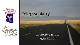
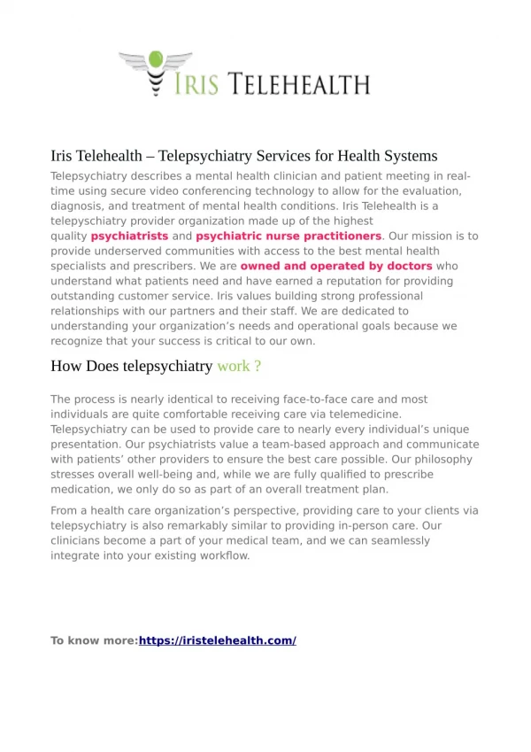
Telepsychiatry • In a sense, telepsychiatry is the “native application” for interactive-video telemedicine. Mental health interactions are straightforward, and demand little other than that the participants see and talk with one another. There is no need for sophisticated peripheral devices such as video oto-ophthalmoscopes or electronic stethoscopes. Perhaps for this reason, telepsychiatry was the first use of interactive video for medical purposes; there are reports dating back to the 1960s and 1970s. • Although telepsychiatry can save patients (and their doctors) the trouble of travelling and also allow some “inhibited” or shy patients to talk more freely, it will always suffer from the lack of the human dimension of direct face-to-face contact.
Teledermatology and Telepsychiatry Part 2 A Practical Guide to Teledermatology Imaging and Evaluation Partly adapted from: Vidmar DA. The Idiot's Guide To Teledermatology Imaging. Department of Military and Emergency Medicine Uniformed Services University of the Health Sciences, Bethesda, US - URI: http://navmedinfo.med.navy.mil/ntbo/TeleDermatology/Idiot's Guide to Tderm Imaging.html
Minimum Imaging and Lighting Equipment Requirements • Light sources (camera’s integral flash unit, Surgical lights, Gooseneck lights, Penlights etc.). • Digital camera capable of at least 600X800 lines per image in “millions of colours”. • Lens capable of taking both longer distance views as well as close ups (i.e. 50-90 mm macro equivalent). A “fixed focus” type lens is not appropriate. • Intrinsic flash which can be set to “auto” or “off”. The Nova Exam light provides maximum illumination, ease of movement, stability and style in a newly designed, attractive small exam light. The unique tilt-and-glide compact base requires minimal floor space. Features: 2600 footcandles, 3100°K color temperature, 20 watt halogen (3,000 hour life), gooseneck with 90° swivel, 40" upright stand plus an additional 28" pole with vertical adjustment and gooseneck.
Know Your Gear • Be sure that you know your camera and flash’s functions and capability. Know your equipment! Be familiar with the manuals. In particular you need to make sure that: • >Your system (camera and flash if included) has sufficient battery power or external power. • >There is enough hard disk card storage remaining on the camera to complete the imaging session with your patient (some digital cameras can write to a built-in ordinary 3.5” floppy diskette). • >Good lighting tips:Intrinsic flash is used most of the time for close ups (8-10 inches). Use obliquely oriented external lighting as primary source for close ups or as supplemental lighting for distance shots (e.g., full body views). ^ Sony Mavica Digital Camera stores photos on a 3.5” floppy
Sony Mavica Digital Camera Sony Mavica Digital Camera stores digital photos on a 3.5” floppy diskette
Identification of Images - Clinical History • Each patient’s images are a matter of clinical record and require ironclad identification. Begin each patient’s imaging session with an identification photo that includes the patient’s name (e.g., John A Smith), date of imaging session, some reliable patient serial number, e.g., NHS number. • Patient identification and demographics are required for medico-legal reasons and for the administrative follow up of the patient. At times, they can even provide some useful clues for the teledermatology consultant.Remember to ask about any medications used by the patient (including over the counter drugs), as well as any drug allergies. • Clinical history: You should have adequate clinical information about the skin condition before you image the patient. This information should be made available to the specialist dermatologist together with the photos(in store-and-forward teledermatology; in real-time teledermatology, the consultant can directly ask the patient).
Background and Positioning • Have your patient remove all medications, make-up, wristwatches, distracting objects or any clothing that interferes or distracts with the taking of the images. Be sure to have a “standby” in attendance when imaging “sensitive” areas (e.g., breasts and genitalia). • Position your patient in front of a reasonably clean, plain background such as a large grey surgical drape or bedsheet. Make sure that the background is neither too bright nor shiny, nor too dark either. Use a patient-to-camera distance that allows the inclusion of all of the appropriate areas. Make sure that your patient is comfortable and that the room is warm enough! • N.B.: Images must not be used for any other purpose (e.g., published in a research paper or an educational atlas) without a separate patient consent.
Shade and Hue Reference • Try to include either a reference colour bar (e.g., MacBeth Colour bar) or a greyscale reference card (see illustration below) in the images. The patient can hold it for you as you take the images. If not available, be sure to include in the viewfinder any very black object (e.g., black computer disk) and any very white object (e.g., a sheet of white bond paper). MacBeth Colour bar >
Areas to Image • This is actually not as difficult as it may seem. You need to do the same thing as any healthcare provider looking at a patient with a skin condition. Resist the urge to immediately get very close to the condition. Be methodical in your approach. Start from a longer distance and then get as close and detailed as required for a particular condition. • Not every patient requires multiple views with detailed lighting techniques. However, it is important to figure out the sorts of views and level of detail that each patient needs.
Helping the Dermatologist Determine the Size of a Lesion • Sometimes, you need to include a ruler in your view to help the dermatologist determine the actual size of a lesion. This becomes very important when following-up the same lesion over time. Superficial Spreading Melanoma, Level IV (Biopsy) - Site: Right Shoulder
Telepathology in the Context of Teledermatology Benign Lymphangio-endothelioma - Site: Right Upper Anterior Thigh Lesion • Dermatologists sometimes rely on microscopic examination of skin biopsy to reach a final diagnosis (dermatopathology). • In a telemedicine setting, the same techniques used in telepathology (tele-histopathology) will generally apply.
Clinical Example - 1 Evaluating a Specific Spot or Solitary Area of Interest e.g., mole on a back or rash on foot. You should include the following views: • Consider taking a view including the whole back for orientation (usually 8 feet). This is frequently not necessary if the skin problem is very well localised. Use the following suggested lighting techniques if you do not have an intrinsic flash.
Clinical Example - 1 (Cont’d) • A medium view (usually 3-5 feet) including some anatomical landmarks for orientation. • At least one close up view in which the lesion occupies about one quarter of the viewfinder (usually 1 foot). • Keep your camera directly perpendicular to what you are trying to photograph.
Clinical Example - 1 (Cont’d) • If your camera’s light source causes glare with close up views, use an oblique source of light positioned at a 45 degrees angle to the area you are trying to image.
Clinical Example - 2 Imaging a Skin Condition Involving Multiple Sites (usually widespread rash). You need the following views: • Long distance views (front and back) at 8-10 feet of distance to illustrate the total area of involvement • A medium distance view to show the worst areas of the rash. Remember to image the areas of the rash that the patient says are the most bothersome. • Also remember to ask about any involvement of palms, soles and genitalia.
Clinical Example - 2 (Cont’d) • If one hand is affected, image the other one for comparison and check the soles as well to see if they also are involved. If they are, image them or at least examine them and make sure that they are not affected. The same thing applies if one foot is involved. • If one elbow is affected, image the other one for comparison and check the knees as well to see if they are involved. If they are, image them or at least examine them and make sure that they are not affected. The same thing applies if a knee is involved.
Clinical Example - 2 (Cont’d) • Close up views of the worst areas of the rash, preferably including areas that are minimally scratched by the patient. • Use a light held at a 45-degree angle to the surface of the skin to bring out the details of topography and surface texture of the involved skin. The lighting technique employed can tremendously affect the image that you are taking. You should be concerned about highlighting texture (e.g., rough, smooth, scaly, etc.) and topography (e.g., flat macule or raised papule or plaque, etc.) for your consultant as well as being concerned about over and under illumination of the skin. A poorly taken image can fool the consultant.
Types of Clinical Images • Overview; long distance view(s) taken to illustrate the distribution of a widespread or multi-focal skin problem. • Regional; medium distance views designed to illustrate the worst or most representative sites or in the case of a skin growth, to provide some orientation as to where it is on the body. • Close Up; very detailed views designed to show the surface texture, topography, colour and architecture of a skin growth or the detailed appearance of a “rash”. As mentioned before, the use of oblique lighting and focus are crucial (and sometime difficult) for this type of image.
On Autofocus • While many digital cameras and other telemedicine imaging devices come equipped with an autofocusing feature, one should not blindly trust them. Make sure that the area of interest is in the dead centre of the viewfinder when the Autofocus feature is engaged, otherwise it will automatically focus on an object that you may not be interested in.
Teledermatology Is It a Cost-Effective Method? • Rueda MF, Kress D, Sidique Y, Barnes B and Jegasothy BV. Teledermatology: Is It a Cost-Effective Method of Dermatologic Consultation? A Preliminary Study. Department of Dermatology, University of Pittsburgh School of Medicine/UPMC Presbyterian, Pittsburgh, PA, US. 2000URI: http://www.upmc.edu/dermatology/ResearchClinical/teledermatology.htm • This study compared two methods of providing dermatological care; a remote consultation using a video camera (teledermatology) and the traditional “face-to-face” visit. The study also examined cost-effectiveness and quality of care provided.
Teledermatology Is It a Cost-Effective Method? (Cont’d) • Having a face-to-face consultation with a dermatologist is the “gold standard” of dermatological care. As a quality control during the study, the authors wanted to compare the gold standard with the teledermatological method performed by another dermatologist to establish if teledermatology is a reliable, reproducible, generalisable, and cost-effective procedure. • If the results of teledermatology rival those of face-to-face consultation, teledermatology may provide a means of increasing access to dermatological care in rural and underserved areas. Patients in these areas can receive care without having to travel long distances and be absent from daily activities. This significant improvement in care may be possible without increasing patients’ or healthcare provider’s expenses.
Teledermatology Is It a Cost-Effective Method? (Cont’d) Equipment • Camera: Panasonic S-VHS Reporter, movie camera 456 Proline • Software: VTEL Smart Station 5.02-System. (Digital Visual Communications) • PC: Compaq Deskpro, Pentium II • Screen: Compaq S900, 1600 x 1200 pixel, non-interlaced, 18 x 18 x 18.5 inches • Speakers: Yamaha YST-M15
Teledermatology Is It a Cost-Effective Method? (Cont’d) Results, Comments and Conclusions • Twenty-nine of 30 diagnoses were concordant between both dermatologists (teledermatologist and another face-to-face dermatologist examined each case). This corresponds to an overall diagnostic accuracy of 96.5%. Study data were not enough to state if the reason for the non-concordant case is related to the method of consultation or to other variables, such as characteristics of the disease or dermatologists’ own experiences. It is not possible to conclude if the discrepancy will be repeated if another two dermatologists observe the case using a face-to-face method.
Teledermatology Is It a Cost-Effective Method? (Cont’d) Results, Comments and Conclusions • Even though the colour assessment was satisfactory in the results, doctors stated the importance of not using fluorescent light to decrease the greenish hue produced by it. Additionally it was observed that Manual Mode instead of Autofocus is better to ensure accuracy. • Light needs and sources depend on individual settings and must be tested before starting the actual consultation process.
Teledermatology Is It a Cost-Effective Method? (Cont’d) Results, Comments and Conclusions • Expertise in operating the equipment to show oral and nasal mucosae should be achieved before the actual consultation process. One patient in the study was not evaluated by the teledermatology method due to lack of expertise of the operator in illuminating and showing oral lesions. Patient was diagnosed by face-to-face method as aphthae. • No patients presented with nodular or deep lesions; thus, the efficacy of the method for these sorts of cases was also not evaluated.
Teledermatology Is It a Cost-Effective Method? (Cont’d) Results, Comments and Conclusions • On Questions about Convenience, it was observed that a great number of patients have to spend a long time travelling to reach the dermatology office. This amount of time and inconvenience will be more than doubled if the visit time and returning time are added. • The method proves to be especially convenient for those patients having to travel long distances to visit the dermatologist.
Teledermatology Is It a Cost-Effective Method? (Cont’d) Results, Comments and Conclusions • On Questions about Quality of Care, the consensus opinion was that teledermatology is good and offers high-quality care; however, the investigational setting must be made more comfortable. 88.2% of patients thought that the quality of care provided by the method was excellent. • Subjective Opinion about the New Method: more than half the patients liked the new method of consultation. The majority of the patients who did not like it were those who do not need it because their travel time is less than one hour. Only two patients from those who did not like the method had to travel more than 1 hour to the visit.