Heart Physiology
Heart Physiology. What a job!. It pushes your 6L of blood through your blood vessels over 1000 times a day!. Cardiac Muscle. Intercalated disks – link every cardiac muscle cell together; composed of Gap Junctions – allow the ions to flow between cells

Heart Physiology
E N D
Presentation Transcript
What a job! • It pushes your 6L of blood through your blood vessels over 1000 times a day!
Cardiac Muscle Intercalated disks– link every cardiac muscle cell together; composed of • Gap Junctions – allow the ions to flow between cells • Desmosomes – physically connect adjacent cells
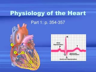
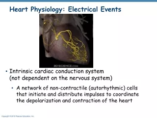
Heart Rhythm • Intrinsic Conduction System (ICS) – “specialized” tissue built into the heart wall; sets basic rhythm, ~75 beats/minute • Extrinsic Control – nervous system can increase or decrease heart rate +
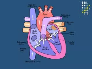
Intrinsic Conduction System Autorhythmic Cells • S-A Node (Sinoatrial node) –located in the wall of the R.A. by the Superior vena cava. • Starts each heartbeat, aka.. “The Pacemaker” • Causes the atria to contract
A-V Node (Atrioventricular node) –located between the atria and ventricles • Contracts when receives impulses from the S-A node • Causes ventricular contraction The signal then travels through the rest of the system: • Bundle of His • Bundle Branches • Purkinjie fibers
4 3 5
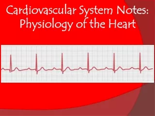
Electrocardiogram [ECG] • Traces the flow of current through the heart
Irregular Heartbeats • Tachycardia – rapid heart rate (>100 beats/min) • Bradycardia– low heart rate (<60 beats/min) • Ischemia – lack of adequate blood supply to heart muscle can lead to • Fibrillation – rapid uncoordinated shuddering of heart muscle (a major cause of death from adult heart attacks)
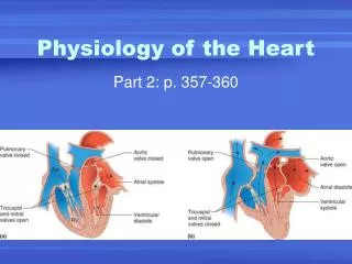
Cardiac Cycle All the events in 1 complete heartbeat (both atria and ventricles contract and relax). Systole – contraction of the ventricles Diastole – relaxation of the ventricles • Events occur in 3 phases
1. Mid-to-Late Diastole • Heart in complete relaxation • Low pressure • Blood flowing into atria and ventricles • Semi-lunar valves closed • A-V valves open END – atria contract and force remaining blood into ventricles
2. Ventricular Systole • Ventricles contract • V pressure increases • A-V valves close • Atria begin filling with blood END – when pressure in V’s is greater than pressure in arteries, semi-lunar valves are forced open • Blood rushes out of V’s
3. Early Diastole • Ventricles relax • Semi-lunar valves close • Ventricular pressure drops END – when pressure in V’s is less than pressure in atria (which have been filling with blood), the A-V valves are forced open • Blood rushes into V’s
Cardiac Output • Stroke Volume (SV) – Volume of blood pumped by each ventricle with each heartbeat • Cardiac Output (CO) – amount of blood pumped by each ventricle in 1 minute = Heart Rate (HR) * Stroke Volume (SV) Normal Adult HR is ~75 beats/minute Average resting SV is ~70 ml/beat What is the average adult CO?