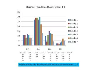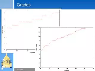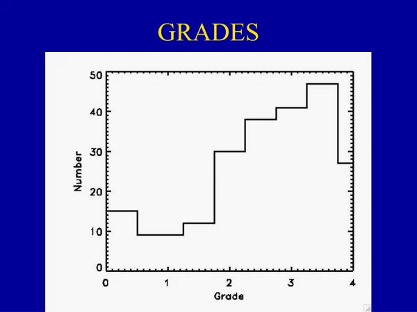
Class grades
3 Quizzes 9/27: Ventricular System 11/20: Brainstem and Basal Ganglia 12/6: Cranial Nerves Clinical Notebooks Due : 11/13 ---- no late submissions accepted. 2 Exams 11/1: Somatosensory System, Visual System, Central Auditory System, and Vestibular System

Class grades
E N D
Presentation Transcript
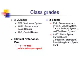
3 Quizzes 9/27: Ventricular System 11/20: Brainstem and Basal Ganglia 12/6: Cranial Nerves Clinical Notebooks Due: 11/13----no late submissions accepted 2 Exams 11/1: Somatosensory System, Visual System, Central Auditory System, and Vestibular System 11/27: Motor System: Cortical Level, Cerebellum, Brainstem, Basal Ganglia and Spinal Cord Class grades
Learning Objectives • 1. Describe the meninges, their locations, and their functions. • 2. Identify parts of the ventricular cavities. • 3. Discuss functions of cerebrospinal fluid. • 4. Describe the mechanism of cerebrospinal fluid production. • 5. Describe the circulation of cerebrospinal fluid. • 6. Explain the diagnostic significance of the cerebrospinal fluid.
Protection of the CNS • Function: • CNS is fairly soft and gelatinous in nature • Basic protection
Meninges of the Brain • Three Meninges:
The Meninges • Bhatnagar & Andy, 1995, Figure 2.45A
Meningeal Layers: Dura Mater • Location: • Function:
Meningeal Layers: Dura Mater • Structure: • Two spaces • Two fibrous layers of the dura
The Meninges • Bhatnagar & Andy, 1995, Figure 2.45A
The Meninges • Love & Webb, 1996, Figure 3-3
Dural Extensions • Falx Cerebri • Location: • Cavity Formations:
Dural Extensions on Midsagittal Section • Bhatnagar & Andy, 1995, Figure 2.43
Dural Extensions on Coronal View • Bhatnagar & Andy, 1995, Figure 2.44A
Dural Extensions • Tentorium Cerebelli • Location: • Tentorial Notch
Dural Extensions on Midsagittal Section • Bhatnagar & Andy, 1995, Figure 2.43
Dural Extensions on Coronal View • Bhatnagar & Andy, 1995, Figure 2.44A
Dural Extensions • Falx Cerebelli • Location:
Dural Extensions on Midsagittal Section • Bhatnagar & Andy, 1995, Figure 2.43
Dural Extensions on Coronal View • Bhatnagar & Andy, 1995, Figure 2.44A
Meningeal Layers: Arachnoid Membrane • Structure: • Location:
Meningeal Layers: Arachnoid Membrane • Spaces: • 1. Subarachnoid space • 2. Subdural space:
The Meninges • Bhatnagar & Andy, 1995, Figure 2.45A
Arachnoid Villi or Granulations • Bhatnagar & Andy, 1995, Figure 2.41
Meningeal Layers: Pia Mater • Location: • Structure:
The Meninges and the Spinal Cord • Similar Structures
The Spinal Cord and Its Meninges • Bhatnagar & Andy, 1995, Figure 2.46
The Ventricular System • Three Parts: • Function:
Ventricular System in Relation to Brain: Lateral View • Bhatnagar & Andy, 1995, Figure 2.37
Ventricular System: Lateral View • Bhatnagar & Andy, 1995, Figure 2.35A
Ventricular System: Dorsal View • Bhatnagar & Andy, 1995, Figure 2.35B
The Lateral Ventricles • Structure and Shape: • Location: • Connection: • Choroid Plexus:
Ventricular System in Relation to Brain: Lateral View • Bhatnagar & Andy, 1995, Figure 2.37
The Third Ventricle • Location and Shape: • Connection: • Choroid Plexus:
Ventricular System in Relation to Brain: Lateral View • Bhatnagar & Andy, 1995, Figure 2.37
The Fourth Ventricle • Location: • Shape: • Structure: • Function:
Ventricular System in Relation to Brain: Lateral View • Bhatnagar & Andy, 1995, Figure 2.37
Subarachnoid Space • Location: • Arachnoid Trabeculae
The Meninges • Bhatnagar & Andy, 1995, Figure 2.45A
Ventricles • Inner Walls
Cerebrospinal Fluid • Structure: • Circulation • Function:
Choroid Plexus • Function: • Location:
Path of CSF Circulation • Pathway: • Flows from the lateral ventricles into the third ventricle • Via Monro’s foramen • Then flows from the third ventricle to the fourth ventricle through the cerebral aqueduct • Then flows from the fourth ventricle into the subarachnoid space through three apertures • Two lateral Foramina of Luschka • One mediodorsal Magendie’s foramen • Then travels to reach the inferior surface of the cerebrum and moves superiorly over the lateral aspect of each hemisphere • Some of it moves into the subarachnoid space around the spinal cord
The Ventricular System: Midsagittal View • Bhatnagar & Andy, 1995, Figure 18.2
Circulation of the CSF • Love & Webb, 1996, Figure 3-6
Clinical Considerations • Drainage of the CSF • Inadequate Drainage of the CSF
Clinical Considerations • Rate of CSF Production • Disassociation between Production and Absorption Rate of the CSF
Clinical Considerations • Hydrocephalus • Increased Pressure in the Brain • Sustained Pressure
MRI of Enlarged Lateral Ventricles Secondary to Hydrocephalus • Bhatnagar & Andy, 1995, Figure 18.3