Download
1 / 10
100 likes | 232 Views
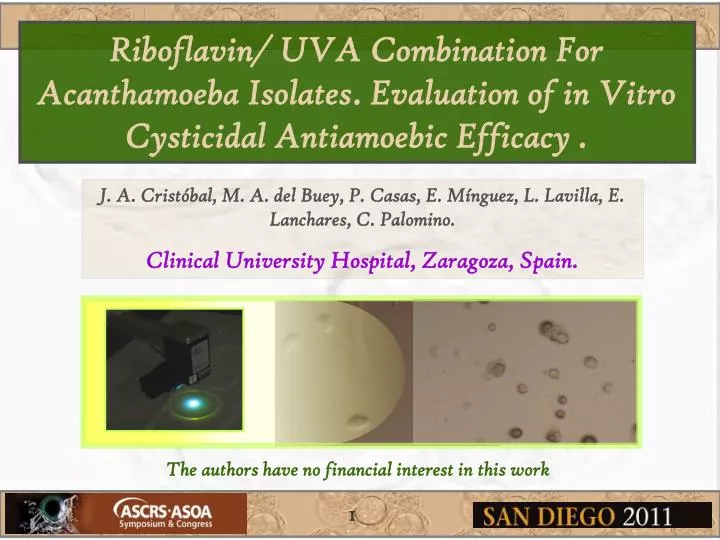
Riboflavin/ UVA Combination For Acanthamoeba Isolates . Evaluation of in Vitro Cysticidal Antiamoebic Efficacy . J. A. Cristóbal, M. A. del Buey, P. Casas, E. Mínguez, L. Lavilla, E. Lanchares, C. Palomino. Clinical University Hospital, Zaragoza, Spain.

E N D
Riboflavin/ UVA Combination For Acanthamoeba Isolates. Evaluation of in Vitro Cysticidal Antiamoebic Efficacy . J. A. Cristóbal, M. A. del Buey, P. Casas, E. Mínguez, L. Lavilla, E. Lanchares, C. Palomino. • Clinical University Hospital, Zaragoza, Spain. The authors have no financial interest in this work 1
INTRODUCTION Acanthamoeba spp.has two morphological forms in its life cycle, an amoeba or trophozoite stage, that feeds and divides, and a resistant cyst stage, that protects the amoeba from adverse environmental conditions. Cysts ofAcanthamoeba.spp: The cyst is the resistance form, can be polygonal shaped and shows a double wall and a nucleus. Trophozoite of Acanthamoeba spp: The trophozoite is the mobile form, nucleus (N), contractile vacuole (vc) and acanthopodia (A) can be seen in the picture above. Acanthamoeba Keratitis (AK) has always presented a medical challenge for most ophthalmologists because it is a frustrating and often devastating infection. Recent in vitro studies have shown an antimicrobial effect of riboflavin and ultraviolet-A (UVA) collagen cross-linking (CXL) on a number of bacterial and fungal pathogens, but their effectiveness in vitro against protozoa has not been proven in published studies. 2
CBM-X-LINKER, irradiance of 3 mW/cm2 and wavelength of 370 nm. The aim of this work is to evaluate the in vitro viability of two different strains of acanthamoeba after UVA-CLX application. We designed a method of assessing the growth of the amoebae in a solid medium, which allows us to reproduce the conditions of in vivo treatment. METHODS Two different strains of Acanthamoeba were identically tested. • Acanthamoeba sp 65 was used as an environmental amoeba isolated from superficial water. • Acanthamoeba sp 7376was used as a clinical strain isolated via corneal scraping for a patient with keratitis. Four groups of treatment were considered: • GROUP 1: 0.1% riboflavin, 30-minute UVA irradiation. • GROUP 2: 0.1% riboflavin, 60-minute UVA irradiation. • GROUP 3 (Control): no riboflavin, no UVA exposition. • GROUP 4: 0.1% riboflavin, no UVA exposition. 3
METHODS The experiments were performed threetimes with each group. The application of UVA was performed with the same parameters used for “in vivo” corneal collagen cross-linking. The diameter of application in all cases exceeded the limits of seeding of protozoa. • The seeding plates for both species of amoebae for each group (including the control group) were evaluated after 24 hours of incubation at 30ºC after the treatment . • If amoebae were observed around the seeding area, the migration radius (from the point of application of the amoeba to the point where trophozoites had migrated) would be measured to quantify the results for the different groups. 4
RESULTS • In all groups and for both isolates studied (environmental and pathogenic strains), cysts and trophozoites were detected at distances from the point of application greater than 5 mm after incubation at 30ºC for 24 hours, indicating the viability of the amoebae tested. • Trophozoites were found at a similar distance from the point of application for all treatment groupsand for the control group for both strains of amoebae (14-15 mm); the only exception was the group exposed to CXL and Riboflavin for 60 min (GROUP 2), for which the distance traveled was slightly lower (12 mm). After 72 hours, the trophozoites had spread across the agar in all groups. • The morphology of the trophozoites at 24 hours after treatment was similar for the control and treatment groups. However, after 60 minutes of CXL application (GROUP 2), the degradation of some cysts was observed. 5
RESULTS A) Point of Acanthamoeba application, diameter less than 5 mm. B) Riboflavin application and UVA radiation, 9 mm of diameter. C) After incubation at 30ºC for 24 hours, Acanthamoeba detection at distances from the application point greater than 5 mm (12-15 mm depending on group). 6
RESULTS A) Untreated cysts of Acanthamoeba 7376 B) Untreated trophozoites of Acanthamoeba 7376. C) Cysts of Acanthamoeba 7376 after treatment with rivoflavine and 60 minutes of Cross-linking. Note the degradation of some cysts with respect to the control. D) Trophozoites of Acanthamoeba 7376 after treatment with rivoflavine and 60 minutes of Cross-linking. Note the similar morphology to untreated control. 7
DISCUSSION • The efficacy of UV light as a disinfectant in contact lens, water or blood concentrates has been investigated in recent years. The eradication of E. Coli with UV-A requires much higher doses and application time than UV-B and UV-C. Nevertheless, UV-B (320-290 nm) and UV-C (290-200 nm) are highly damaging to DNA and are associated with a high risk of causing serious damage to cells and tissues. Thus, they are not used in humans. • Our study was performed using the criteria for the UVA-riboflavin treatment of keratoconus described by Wollensak et al in 2003. Our results are consistent with those presented by Maya et al., who conclude that the total inactivation of amoebae requires higher UV doses than are necessary for bacteria. • We must keep in mind that our study only assesses in vitro antiamoebicidal activity under levels of UVA radiation used in human therapy; it does not assess the effectiveness of the same treatment with biological tissue that exhibits secondary changes such as edema or corneal melting. 8
CONCLUSIONS • In conclusion, given that 30 or 60 minutes of UVA exposure (3 mW/cm2, 370 nm) or riboflavin therapy is not effective against the two different acanthamoeba strains examined in our study (environmental acanthamoeba spp 65 and pathogenic acanthamoeba spp 7376), it seems reasonable to suppose that a longer exposure period could achieve the desired effect. However, such a treatment is not advisable due to toxicity and risk of tissue damage. • In vitro results are not always indicative of in vivo efficacy; thus, future studies should further evaluate this treatment for AK. Further studies using Acanthamoeba cultures on corneas may provide more information. 9
The authors gratefully acknowledge the research support of the Spanish Ministry of Education and Science through the research project DPI2008-02335. REFERENCES • Wollensak G, Spoerl E, Seiler T. Stress-strain measurements of human and porcine corneas after riboflavin-ultraviolet-A-induced cross-linking. J Cataract Refract Surg 2003;29:1780-1785. • Martins SA, Combs JC, Noguera G, et al. Antimicrobial efficacy of riboflavin/UVA combination (365 nm) in vitro for bacterial and fungal isolates: a potential new treatment for infectious keratitis. Invest Ophthalmol Vis Sci 2008;49:3402-3408. • Iseli HP, Thiel MA, Hafezi F, Kampmeier J, Seiler T. Ultraviolet A/riboflavin corneal cross-linking for infectious keratitis associated with corneal melts. Cornea 2008;27:590-594. • Maya C, Beltrán N, Jimenez B, Bonilla P. Evaluation of the UV disinfection process in bacteria and amphizoic amoeba inactivation. Water Sci Tech 2003;3:285-291. • Makdoumi K, Mortensen J, Crafoord S. Infectious keratitis treated with corneal crosslinking. Cornea 2010;29:1353-1358. • Khan YA, Kashiwabuchi RT, Martins SA, et al. Riboflavin and ultraviolet light A therapy as an adjuvant treatment for medically refractive acanthamoeba keratitis report of 3 cases. Ophthalmology 2011;118: 324-31. • Ubomba-Jaswa E, Navntoft C, Polo-López MI, Fernandez-Ibáñez P, McGuigan KG. Solar disinfection of drinking water (SODIS): an investigation of the effect of UV-A dose on inactivation efficiency. Photochem Photobiol Sci 2009;8:587-595 madelbuey@telefonica.net 10