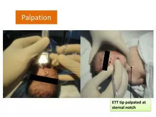
Palpation
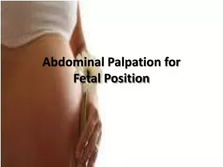
Palpation. Palpation is the most important in examination of abdomen. The preparation of patient: with the patient in supine position, the head should be elevated on a pillow, patient ’ s arms relax at the two sides of the body, flex his thighs and knees, relax his abdominal muscles.

Palpation
E N D
Presentation Transcript
Palpation is the most important in examination of abdomen. The preparation of patient: with the patient in supine position, the head should be elevated on a pillow, patient’s arms relax at the two sides of the body, flex his thighs and knees, relax his abdominal muscles
According to different parts and organs of examination, the patient can be in right lateral decubitus position such as the examination of spleen, or standing position such as the examination of kidney.
The position of doctor the doctor stands at the right side of patient, warm hands, use your palmar aspect of fingers examine gently and lightly from superficial to deep, and from healthy part to lesion area.
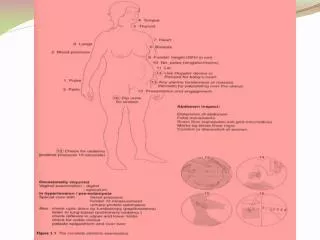
The sequence of palpation usually the sequence of palpation is contraclock direction:left lower left lumber left upper epigastric right upper right lumber right lower hypogastric umbilical.
the palpating methods 1、浅部触诊法 (light palpation)2、深部触诊法 (deep palpation)(1).深部滑行 (deep slipping palpation)(2).双手触诊法 (bimanual palpation)(3).深压触诊法 (deep press palpation)(4).冲击触诊法 (ballottement)
The contents of palpation1.abdominal muscles tensity 2.tenderness and rebound tenderness 3.abdominal organs4.abdominal masses 5.fluid thrill 6.succussion splash
1.abdominal muscle tensity(1). Increased tensity of generalized abdominal muscles Acute diffuse peritonitis caused by gastrointestinal perforation. In perforation, the muscle rigidity is very obvious, the abdominal wall is like board hard, so we call board-like abdomen
Dough kneading sensation TB peritonitis carcinomatous peritonitis metastatic carcinoma of peritonium
(2). Increased tensity of located abdominal muscles one organ inflammation right upper abdomen acute cholecystitis: involved peritoneum right lower abdomen acute appendicitis: involved peritoneum
Decreased tensity of abdominal wall decreased chronic consumptive disease: cachexia after tapping ascites disappeared abdominal muscles paralysis myasthenia gravis spinal cord trauma
2.Tenderness and rebound tendernessusually caused by inflammation, carcinoma and TB. The part of tenderness is usually the location of lesion.for example, tenderness in right upper abdomen: hepatitis cholecystitis.
Lumber region kidney stoneright lower abdomen appendicitis epigastric region peptic ulcer umbilical small intestine diseases ascariasis rebound tenderness when inflammation involve parietal peritoneum such as acute peritonitis, acute appendicitis. the rebound tenderness is positive
3.Palpation of the organs1). palpation of the liver palpating methods palpation with one hand bimanual palpation clasping palpation ballottement
When you palpate the liver you should pay special attention to the following items (1) size (2) consistency (3) contour margin (4) tenderness (5) pulsation (6) friction sound (7) liver thrill
(1). The size of liverin healthy person the liver is not palpable or palpable within 1 cm below the costal margin 3 cm below the xiphoid hepatomegaly diffuse hepatitis fatly liver early cirrhosis of liver hepatic engorgement
located enlargement of liver hepatic cyst hepatic abscess shrinking of liver acute liver necrosis cirrhosis of liver hepatometry midclavicular line how many cm below costal margin abdominal middle line how many cm below xiphoid process
(2)Consistency the consistency of liver is divided into 3 degrees soft as like lips seen in normal liver middle hard as like nose acute chronic hepatitis hard as like frontalis cirrhosis carcinoma fluctuation big surface cyst
(3) Contour and margin normal liver: the surface is smooth and margin is regular irregular nodular dull: cancer (4) tenderness normal liver: no tenderness light: hepatitis sever: hepatic abscess
(5) Pulsationnormal liver: no pulsation distensible pulsation: tricuspid valve incompetencetransmitted pulsation: aneurysm (6) friction sound of the hepatic area: perihepatitis (7) liver thrill: echinococcosis
The error of palpation(1) patient can`t coordinate with doctor (2) massive liver to palpate over liver (3) the doctor`s hand presses too heavy to move liver down
(4) some organs may be misapprehended the liver such as: transverse colon lower extreme of right kidney tendon of abdominal rectus
2). Palpation of spleenthe position of the patient supine right lateral decubitus palpating methods palpation with single hand bimanual palpation ballottement
right lateral decubitus position bimanual palpation
Splenometry1line (A-B line) midclavicular line 2line (A-C line) the longest line 3 line (D-F line)
Splenomegalylight <2 cm chronic hepatitis, typhoid fever, middle 3 cm – umbilicus cirrhosis of liver chronic hemolytic jaundice heavy below umbilicus chronic granulocytic leukemia myelofibrosis
Some organs may be misapprehend the spleen (1) enlargement of left kidney lower extreme - dull edge (2)enlargement of left lobe of liver no notch (3)cyst of pancreatic trailno notch
3).Palpation of the gallbladder Murphy’s sign: acute cholecystitis courvoisier’s sign pacreatic carcinoma
4). Palpation of kidneybimanual palpation to palpate right kidney
normal: not palpable palpable: (1) nephroptosis >1\2kindey palpable smooth surface middle hard tenderness (-) (2)wandering kidney (3) enlargement of kidney hydronephrosis pyonephrosis tumor
Tenderness pointskidney urinary tube point (1) upper ureter point (2) middle ureter point ureteritis ureterolithiasis (3) costovertebral(4) costolumber pyelonephritis TB kidney pyelolithiasis
Normal: empty not palpable distended: palpable round fluid-filled smooth disappear after urination seen in unconsciousness, after anesthesia, retention of urine
Normal: not palpable epigastric tenderness: acute pancreatitis epigastric cystic mass, under the liver, no movement, smooth, no tenderness pancreatic pseudocyst