Download
1 / 16
170 likes | 191 Views
Discover the composition and behavior of the plasma membrane through experiments and models, showcasing its crucial role in cell biology.

E N D
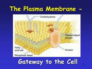
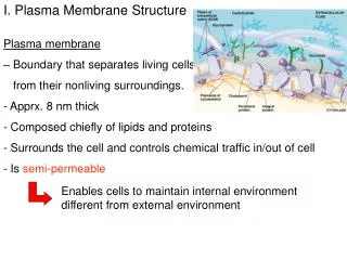
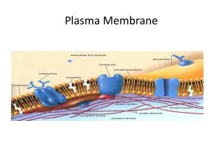
Figure 7.1 • Overview: Life at the Edge • The plasma membrane • Is the boundary that separates the living cell from its nonliving surroundings • The plasma membrane exhibits selective permeability • It allows some substances to cross it more easily than others
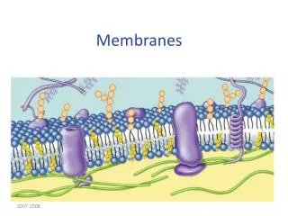
WATER Hydrophilic head Hydrophobic tail WATER Figure 7.2 Membrane Models: Scientific Inquiry • Scientists studying the plasma membrane • Reasoned that it must be a phospholipid bilayer
The Davson-Danielli sandwich model of membrane structure • Stated that the membrane was made up of a phospholipid bilayer sandwiched between two protein layers • Was supported by electron microscope pictures of membranes
Hydrophobic region of protein Phospholipid bilayer Figure 7.3 Hydrophobic region of protein Fluid Mosaic Model • In 1972, Singer and Nicolson • Proposed that membrane proteins are dispersed and individually inserted into the phospholipid bilayer
A cell membrane can be split into its two layers, revealing the ultrastructure of the membrane’s interior. APPLICATION TECHNIQUE Extracellular layer Proteins Knife Plasma membrane Cytoplasmic layer These SEMs show membrane proteins (the “bumps”) in the two layers, demonstrating that proteins are embedded in the phospholipid bilayer. RESULTS • Freeze-fracture studies of the plasma membrane • Supported the fluid mosaic model of membrane structure A cell is frozen and fractured with a knife. The fracture plane often follows the hydrophobic interior of a membrane, splitting the phospholipid bilayer into two separated layers. The membrane proteins go wholly with one of the layers. Figure 7.4 Extracellular layer Cytoplasmic layer
Lateral movement (~107 times per second) Flip-flop (~ once per month) (a) Movement of phospholipids Figure 7.5 A The Fluidity of Membranes • Phospholipids in the plasma membrane • Can move within the bilayer
Membrane proteins EXPERIMENT Researchers labeled the plasma mambrane proteins of a mouse cell and a human cell with two different markers and fused the cells. Using a microscope, they observed the markers on the hybrid cell. RESULTS Mouse cell Mixed proteins after 1 hour Human cell Hybrid cell + CONCLUSION The mixing of the mouse and human membrane proteins indicates that at least some membrane proteins move sideways within the plane of the plasma membrane. Figure 7.6 • Proteins in the plasma membrane • Can drift within the bilayer
Fluid Viscous Unsaturated hydrocarbon tails with kinks Saturated hydro- Carbon tails (b) Membrane fluidity Figure 7.5 B • The type of hydrocarbon tails in phospholipids • Affects the fluidity of the plasma membrane
Cholesterol Figure 7.5 (c) Cholesterol within the animal cell membrane • The steroid cholesterol • Has different effects on membrane fluidity at different temperatures
Glycoprotein Carbohydrate Glycolipid EXTRACELLULAR SIDE OF MEMBRANE Microfilaments of cytoskeleton Peripheral protein Cholesterol Integral protein CYTOPLASMIC SIDE OF MEMBRANE Figure 7.7 Membrane Proteins and Their Functions • A membrane • Is a collage of different proteins embedded in the fluid matrix of the lipid bilayer Fibers of extracellular matrix (ECM)
N-terminus C-terminus CYTOPLASMIC SIDE a Helix Figure 7.8 • Integral proteins • Penetrate the hydrophobic core of the lipid bilayer • Are often transmembrane proteins, completely spanning the membrane EXTRACELLULAR SIDE
Transport. (left) A protein that spans the membrane may provide a hydrophilic channel across the membrane that is selective for a particular solute. (right) Other transport proteins shuttle a substance from one side to the other by changing shape. Some of these proteins hydrolyze ATP as an energy ssource to actively pump substances across the membrane. (a) ATP (b) Enzymatic activity. A protein built into the membrane may be an enzyme with its active site exposed to substances in the adjacent solution. In some cases, several enzymes in a membrane are organized as a team that carries out sequential steps of a metabolic pathway. Enzymes Signal transduction. A membrane protein may have a binding site with a specific shape that fits the shape of a chemical messenger, such as a hormone. The external messenger (signal) may cause a conformational change in the protein (receptor) that relays the message to the inside of the cell. (c) Signal Receptor Figure 7.9 • An overview of six major functions of membrane proteins
(d) Cell-cell recognition. Some glyco-proteins serve as identification tags that are specifically recognized by other cells. Glyco- protein (e) Intercellular joining. Membrane proteins of adjacent cells may hook together in various kinds of junctions, such as gap junctions or tight junctions (see Figure 6.31). (f) Attachment to the cytoskeleton and extracellular matrix (ECM). Microfilaments or other elements of the cytoskeleton may be bonded to membrane proteins, a function that helps maintain cell shape and stabilizes the location of certain membrane proteins. Proteins that adhere to the ECM can coordinate extracellular and intracellular changes (see Figure 6.29). Figure 7.9
1 Transmembrane glycoproteins Secretory protein Glycolipid 2 Golgi apparatus Vesicle 3 Plasma membrane: Cytoplasmic face 4 Extracellular face Transmembrane glycoprotein Secreted protein Membrane glycolipid Figure 7.10 Synthesis and Sidedness of Membranes • Membrane proteins and lipids • Are synthesized in the ER and Golgi apparatus ER
Concept 7.2: Membrane structure results in selective permeability • A cell must exchange materials with its surroundings, a process controlled by the plasma membrane