Download
1 / 15
270 likes | 1.39k Views
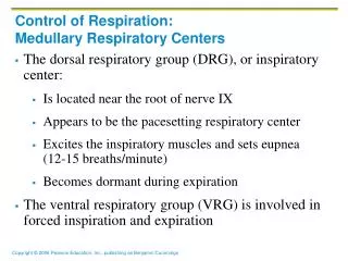
Control of Respiration: Medullary Respiratory Centers. The dorsal respiratory group (DRG), or inspiratory center: Is located near the root of nerve IX Appears to be the pacesetting respiratory center Excites the inspiratory muscles and sets eupnea (12-15 breaths/minute)

E N D
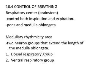
Control of Respiration: Medullary Respiratory Centers • The dorsal respiratory group (DRG), or inspiratory center: • Is located near the root of nerve IX • Appears to be the pacesetting respiratory center • Excites the inspiratory muscles and sets eupnea (12-15 breaths/minute) • Becomes dormant during expiration • The ventral respiratory group (VRG) is involved in forced inspiration and expiration
Control of Respiration: Pons Respiratory Centers • Pons centers: • Influence and modify activity of the medullary centers • Smooth out inspiration and expiration transitions and vice versa • The pontine respiratory group (PRG) – continuously inhibits the inspiration center
Respiratory Rhythm • A result of reciprocal inhibition of the interconnected neuronal networks in the medulla • Other theories include • Inspiratory neurons are pacemakers and have intrinsic automaticity and rhythmicity • Stretch receptors in the lungs establish respiratory rhythm
Depth and Rate of Breathing • Inspiratory depth is determined by how actively the respiratory center stimulates the respiratory muscles • Rate of respiration is determined by how long the inspiratory center is active • Respiratory centers in the pons and medulla are sensitive to both excitatory and inhibitory stimuli
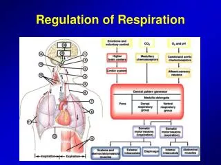
Medullary Respiratory Centers Figure 22.25
Depth and Rate of Breathing: Reflexes • Pulmonary irritant reflexes – irritants promote reflexive constriction of air passages • Inflation reflex (Hering-Breuer) – stretch receptors in the lungs are stimulated by lung inflation • Upon inflation, inhibitory signals are sent to the medullary inspiration center to end inhalation and allow expiration
Depth and Rate of Breathing: Higher Brain Centers • Hypothalamic controls act through the limbic system to modify rate and depth of respiration • Example: breath holding that occurs in anger • A rise in body temperature acts to increase respiratory rate • Cortical controls are direct signals from the cerebral motor cortex that bypass medullary controls • Examples: voluntary breath holding, taking a deep breath
Depth and Rate of Breathing: PCO2 • Changing PCO2 levels are monitored by chemoreceptors of the brain stem • Carbon dioxide in the blood diffuses into the cerebrospinal fluid where it is hydrated • Resulting carbonic acid dissociates, releasing hydrogen ions • PCO2 levels rise (hypercapnia) resulting in increased depth and rate of breathing
Depth and Rate of Breathing: PCO2 • Hyperventilation – increased depth and rate of breathing that: • Quickly flushes carbon dioxide from the blood • Occurs in response to hypercapnia • Though a rise CO2 acts as the original stimulus, control of breathing at rest is regulated by the hydrogen ion concentration in the brain
Depth and Rate of Breathing: PCO2 • Hypoventilation – slow and shallow breathing due to abnormally low PCO2 levels • Apnea (breathing cessation) may occur until PCO2 levels rise
Depth and Rate of Breathing: PCO2 • Arterial oxygen levels are monitored by the aortic and carotid bodies • Substantial drops in arterial PO2 (to 60 mm Hg) are needed before oxygen levels become a major stimulus for increased ventilation • If carbon dioxide is not removed (e.g., as in emphysema and chronic bronchitis), chemoreceptors become unresponsive to PCO2 chemical stimuli • In such cases, PO2 levels become the principal respiratory stimulus (hypoxic drive)
Depth and Rate of Breathing: Arterial pH • Changes in arterial pH can modify respiratory rate even if carbon dioxide and oxygen levels are normal • Increased ventilation in response to falling pH is mediated by peripheral chemoreceptors
Peripheral Chemoreceptors Figure 22.27