Download
1 / 49
540 likes | 707 Views
Hypertrophic Cardiomyopathy (HCM) is a heart condition characterized by inappropriate myocardial hypertrophy, affecting a small percentage of the population. Learn about its pathology, diagnosis, symptoms, and treatment options.

E N D
HYPERTROPHIC CARDIOMYOPATHY Anthony B. King Jr., M.D., F.P.C.C Cardiac Pacing and Electrophysiology
Hypertrophic Cardiomyopathy (HCM) • First described in 1950s • Inappropriate myocardial hypertrophy in absence of obvious cause of LVH • Often the IVS of nondilated LV with hyperdynamic systolic function • If there is dynamic pressure gradient in subaortic area – HOCM (only ¼) • Abnormal stiffness of LV and impaired filling (diastolic dysfunction) – high LVEDP > pulmonary congestion and dyspnea (most common)
HCM • Overall Prevalence is Low • 0.2% (1/500) of general population and in 0.5% of unselected patients referred for an Echo • It may be the most common genetically transmitted cardiac disorder
HCM: Pathology Macroscopic : • Marked increase in myocardial mass • LV cavities are small • LV> RV • Atria dilated, often hypertrophied • Diastolic Dysfunction • Mitral Regurgitation
HCM:Pattern and Extent of LVH • Vary from patient to patient, heterogeneity in amount of LVH in different regions • Most frequent – ASH, disproportionate hypertrophy of septum and anterolateral wall • 30% localized and mild hypertrophy in single region • Some in unusual location (posterior portion of septum, posterobasal free wall and midventricular) • Hypertrophy is dynamic, may be seen in infants but usually develops during adolescence
Apical Cardiomyopathy (ACM) • Variant with predominant involvement of Apex • Common in Japan, ½ of HCM in Japan • Spadelike configuration during LV angio • Giant negative T wave in precordial leads • Absence of gradient, mild symptoms and benign course
HCM: Histology • Myocardial hypertrophy and gross disorganization of muscle bundles – whorled patterns, abnormalities in cell-to-cell arrangement (disarray) • Fibrosis, may be extensive –visible scars • All all HCM have some disarray and most have involvement of 5% or more of myocardium • Abnormal intramural coronary arteries with small lumen size and thickening of vessel wall (.80%) – may be responsible for myocardial ischemia
Histologic section to show the characteristic myofibre disarray in addition to hypertrophy in hypertrophic cardiomyopathy.
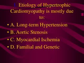
HCM: ETIOLOGY • Autosomal dominant in at least 50% • Sporadic forms – in some if not all are due to spontaneous mutation • Genetic testing not yet easily available and remains largely a research tool
HCM: Pathophysiology • SYSTOLE • DIASTOLE • MYOCARDIAL ISCHEMIA
HCM SYSTOLE: • Dynamic pressure gradient across the LV outflow tract A. Small outflow tract – septal hypertrophy and possible abnormal location of MV B. SAM or systolic anterior motion of MV leaflets against septum (venturi effect)
HCM DIASTOLE • Diastolic dysfunction rest or stress, whether with gradient or none, whether symptomatic or not • Independent of extent and distribution of LVH • Increased filling pressure due to poor LV relaxation and distensability • Fibrosis and cellular disorganization – less distensable
HCM MYOCARDIAL ISCHEMIA • Increased muscle mass • Inadequate capillary density • Elevated diastolic filling pressure • Abnormal intramural coronary arteries • Impaired vasodilatory reserve • Systolic compression of arteries • Enhanced myocardial O2 demand (increased wall stress)
HCM: Symptoms • Very variable • Majority are asymptomatic or just mild • SCD – may be first clinical manifestation • Syncope and SCD in competitive sports • Usually in children, higher mortality in young • In elderly – mild LVH, frequently with outflow gradients and marked symptoms late in life (after 55) • More in men but women more severely disabled
HCM: Symptoms • Dyspnea – increased pulm venous pressure • Angina (75%) • Fatigue, presyncope, syncope – common • Palpitations, PNC, overt CHF and dizziness – less common • Severe CHF culminating in Death • Exacerbated by exertion
ANGINA • 20% have concurrent CAD • AMI may occur in absence of narrowing of extramural arteries SYNCOPE • Inadequate CO – outflow gradient or ischemia • Arrhythmias – SVT (AF) or VT • In young with VT on Holter – increased risk for SCD • In elderly – not an ominous finding
HCM: PE • May be normal in asymptomatic w/o gradient (ACM) • LV lift and S4 • Displaced apex beat, forceful and diffuse • Prominent A wave • Harsh systolic murmur, crescendo-decrescendo, b/w apex and left sternum • Holosystolic if with MR • Murmur augmented by Valsalva, standing, exercise, nitrates
HCM: ECG • Usually abnormal • Normal in 15-25% (localized LVH) • ST and T abnormalities with LVH – most common • QRS complex tallest in midprecordium • Prominent Q waves (20-50%) – inferior and precordial (mimic AMI) • Abnormal axis, P wave abnormality • WPW – uncommon • Abnormal AV conduction (BBB, AVB)
HCM: ECHOCARDIOGRAPHY • Most widely used in evaluation of HCM • Useful in suspected HCM and screening of relatives • Useful in identifying and quantifying morphologic features (distribution of septal hypertrophy), functional aspects (hypercontractile state), hemodynamic findings (outflow gradient)
HCM: Radionuclide Scanning • Reversible thallium defects, indicative of ischemia, common in HCM in absence of obstructive CAD • Seen in young patient with history of syncope and SCD- probable mechanism of demise in this patients
HCM: Cardiac Catheterization • Not required for diagnosis • Reserved when CAD considered or when invasive modalities considered (DDD pacing, surgery) • Phasic narrowing and assoc abnormalities of flow during systole in LAD and septal perforators • LV angio – spadelike deformity, virtual obliteration of cavity at end systole • Determine amount of outflow gradient
HCM: Arrhythmias and Holter • Most common cause of SCD • Poorly tolerated due to systolic and diastolic dysfunction • VA common (>3/4) on Holter • NSVT – ¼ , SuVT – uncommon • Overall predictive value is limited • SupraVT ¼ to ½. AF (10%)
SAECG, HRV – less useful in risk stratification EPS – role in identifying high risk for SCD is controversial and possibly limited value Tilt Table Testing – limited value CXR – findings are variable, from normal to marked cardiomegaly
HCM: Natural History • In many asymptomatic or mild which improve in 5-10 years • Annual mortality –3% in large referral centers, 1% when all patients included • SCD higher in children –6%/year • Clinical deterioration – SLOW • % of severely symptomatic inc with AGE
HCM: Natural History • Onset of AF – increase in symptoms • Progression to LV Dilatation (DCM) 10-15% - those with marked septal hypertrophy and has poor prognosis • Due to wall thinning and scarring from ISCHEMIA • In some children, findings of HCM on Echo may develop despite a previous normal Echo (not common in Adults)
HCM:SCD “Death is most often sudden in HCM and may occur in previously asymptomatic patients, in individuals who were unaware they had the disease, and in patients with otherwise stable course.” • Most common abnormality found in autopsy in young competitive athletes with SCD.
HCM: SUDDEN CARDIAC DEATH (SCD) • HIGH RISK • Young age (<30) at diagnosis • FH of HCM with SCD (malignant FH) • Abnormal BP response to exercise • Genetic abnormalities assoc with inc SCD • Hx of Syncope in children • NSVT on 48 Hour Holter • Massive LVH 8. Brady and disease of AV conduction may play a role
HCM: Management • Alleviation of symptoms, prevent complications and reduction in death. • No data on asymptomatic patients • Avoid digitalis unless with AF or CHF • Cautious diuretic therapy • Mainstay: Bblockers, alternate: CCB or Both • Vast majority – only medical, 5-10% require invasive intervention • Others: Disopyramide, Amio, Sotalol • Anticoagulation in AF, Endocarditis Prophylaxis
HCM: Management INTERVENTIONS: • DDD Pacemaker • Septal Ablation • Surgical – Myectomy, MV Replacement • AICD • Cardiac Transplantation
HCM: Indications for Pacing (ACC/AHA Guidelines) Class I – for sinus node dysfunction or AVB Class II a – None Class II b 1. Medically refractory, symptomatic HCM with significant resting or provoked LV outflow tract obstruction (evid: C) Class III 1. Asymptomatic or medically controlled 2. Symptomatic but w/o evid of LV outflow obstruction