Download
1 / 20
200 likes | 408 Views
Optical Tweezers status and biophysical applications. Lene Oddershede, Lektor, PhD, Niels Bohr Institutet Københavns Universitet. Nano Toolbox. Atomic force microscopy (nano-Newton, nano-m): Unfolding of proteins, binding strengths Optical tweezers (pico-Newton, nano-m):

E N D
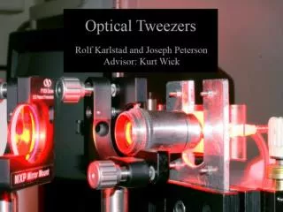
Optical Tweezersstatus and biophysical applications Lene Oddershede, Lektor, PhD, Niels Bohr Institutet Københavns Universitet
Nano Toolbox Atomic force microscopy (nano-Newton, nano-m): Unfolding of proteins, binding strengths Optical tweezers (pico-Newton, nano-m): Single molecule motors, biopolymers Fluorescense (nano-m toÅngstrøm): Single molekyle reaktions, visualization Genetic manipulation Elektron mikroscopy Future use: ’drug delivery’, nano-robots, nano-elektronics ...
Techical know-how 2 force-scope optical tweezers: 3D spatial resolution ~1nm Temporal resolution ~ MHz Single particle tracking: 3D spatial resolution ~5nm Temporal resolution ~ 25 Hz Micropipettes Fluorescens microscopy Genetic manipulations Oddershede, Grego, Nørrelykke, Berg-Sørensen, Probe Microscopy vol. 2, p129 (2001) K. Berg-Sørensen and H. Flyvbjerg, Rev. Sci. Ins. 75, p.594 (2004) Dreyer, Berg-Sørensen, Oddershede, Applied Optics vol. 43, p1991 (2004)
Characteristic photodiode powerloss Silicon is transparant to infrared light. Makes diode a 1st order filter. 8 kHzand depends on laser intensity. a: fraction of incoming power that is correctly detected in depletion layer. Berg-Sørensen, Oddershede, Florin, and Flyvbjerg, J.Appl. Physics. vol. 93 p.3167 (2003)
What can be trapped? Gold nano-particles Succesful optical trapping of 18 nm - 254nm gold particles Literature: 36.4 nm and 40 nm trapped in 3D Mie particles could not be trapped in 3D P.M. Hansen, V.K. Bhatia, N. Harrit, L. Oddershede, Nano Letters, vol.5, p.1937 (2005)
Gold nano rods – nano rotators We have succesfully optically trapped in 3D gold nanorods with diameters ranging from 8nm to 44nm and aspect rations between 1.7 and 5.6 (lenghts up to 85 nm). The rods align with the E-field, can be used as nano-rotators. The optical forces correlate with polarizability of rod. Selhuber-Unkel, Zins, Shubert, Sönnichsen, Oddershede, Nano Letters vol. 8 p.2998 (2008).
Silver nano-particles Change in contrast with size. Has been seen for Au previously, first time for Ag. r: field reflectivity (background) s: scattered field with phase f. the last term is proportional to d3 and dominates for small particles. We optically trap Ag particles with diameters 20nm – 275 nm Spring constants have similar behavior as for Au. Ag nano-particles enhance fluorescense (Au quenche) L. Bosanac, T. Aabo, P.M. Bendix, L.B. Oddershede, Nano Letters vol.8 p.1937 (2008)
Quantum Dots • Quantum dots are the super nova of the nano world. • Very broad absorption band but narrow emission, characterized by blinking. • Superior bleaching properties. • Made of semiconductor material • Ours are of CdSe with a core diameter of ~10 nm, zink-sulfide shell, • and with an emission wavelength of 655 nm. • We have proven possible 3D optical trapping of individual quantum dots. • This makes possible simultaneous manipulation and visualisation. Jauffred, Richardson, Oddershede, Nano Letters vol.8 p.3376 (2008).
Polarizability of an individual Qdot The obtained spring constant, k, carries information about the interaction between the EM field and the Qdot. Using the following relations, the polarization of an individual Qdot can be found: where I : intensity P: total laser power delivered at the sample x,y: directions orthogonal to the light propagation s: width of diffraction limited laser spot. Normalized by vacuum permittivity, we obtain a = 2.8 x 10⁷ Å3. Literature very sparse, only one value reported for Qdots with radius=2nm: a ~ 104 Å3. Jauffred, Richardson, Oddershede, Nano Letters vol.8 p.3376 (2008).
Physiological damage? • It is important to consider the possible physiological damage done by the • optical trap on the trapped cell. • One key issue is to choose a wavelength which is not absorbed by water • (would create heating) and not absorbed by biological specimen either. • Infra-red lasers fulfill these criteria. • To address possible physiological damage by a 1064 nm laser on living • organisms, different bifferent bacterial types were optically trapped. • Simultaneously, the emission from a pH sensitive fluorophore (CFDA, GFP) • inside the organism was monitored. • The capability of a cell to maintain a pH gradient across the cell wall is a • measure of its physiological condition. Healthy cells are able to maintain • a gradient, comprised cells not to the same extend.
Physiological damage of E. coli E. coli expressing GFP Trapped with 6 mW Trapped with 18 mW
GFP, anaerobic (weak signal) Listeria CFDA, aerobic CFDA, aerobic monocytogenes innocua CFDA, anaerobic CFDA, anaerobic
Physiological damage depends on growth conditions and bacterial species M.B. Rasmussen, L. Oddershede, and H. Siegumfeldt, Applied and Environmental Microbiology, vol. 74 p.598 (2008)
Optical tweezers can trap cytoplasmatic organelles without perturbing the cellular membrane
Viscoelasticity of yeast cell cytoplasm • Fedt kugler i levende celler kan benyttes som håndtag for OP • De bevæger sig subdiffusivt pga. microtubulus and aktin netværk • Mere aktin ved cellens ender gør, at lipid kuglerne bevæger sig • mindre frit der.
Brownsk bevægelse: Anormal diffusion: Varians: <x2(t)> = 2Dta Varians: <x2(t)> = 2Dt Normal diffusion = 1 Super diffusion > 1 Subdiffusion 0 < < 1 Begrænset = 0 • Polymer netværk • ( = 0.75) 1. Molekylære motorer 2. Polymerisering 2. Membraner ( = 0.66) 3. Cytoplasmatisk strømning
How do the lipid granules move? mostly bý subdiffusion, a=0.75 Normal diffusion Super diffusion Plateau pga. endelig celle størrelse Optical tweezers Single particle tracking
Nano-mekanics of cell division S. pombe strains expressing green fluorescent protein (GFP): IDEA: Wish to manipulate organelles expressing GFP and use nano particles as handles for the optical tweezers. S. pombe perfect model system.
Mikroinjection of nano partikler Problem: S. pombe has an extremely stiff cell wall. Solution: Enzymatic breakdown of outer parts, microinjection of protoplast, followed by cell regeneration.
Conclusions - Perspectives • Technical issues • Possible to trap individual nano-particles, e.g. gold and silver spheres, gold • rods, • Non-invasive, provided correct wavelenght, small energy deposit. • Examples of biological applications • Molecular motors: polymerase, kinesin, ribosome, virus. • Mechanical strength of mRNA hairpins, pseudoknuder, • Non-equilibrium nano-scale systems. • In vivo studies, viscoelasticity of cellular cytoplasm. • Nano-mechanics of cell division • Future • Single molecule studies of nanotoxicology • Force measurements in vivo • Viral infections • Combination with other techniques • Check out; www.nbi.dk/~tweezer