Understanding Cardiac Conducting System: Heart Beat and Blood Pressure Control
Explore the origin and regulation of the heart beat, normal and abnormal ECGs, blood pressure measurements, and risks of hypertension. Learn how the autonomic nervous system and hormonal control influence heart rate and blood pressure.

Understanding Cardiac Conducting System: Heart Beat and Blood Pressure Control
E N D
Presentation Transcript
Learning Outcomes The heart beat originates in the heart itself but is regulated by both nervous and hormonal control. The autorhythmic cells of the sinoatrial node (SAN) or pacemaker set the rate at which cardiac muscle cells contract. The timing of cardiac cells contracting is controlled by the impulse from the SAN spreading through the atria and then travelling to the atrioventricular node (AVN) and then through the ventricles. These impulses generate currents that can be detected by an electrocardiogram (ECG). Examine normal and abnormal ECGs. The medulla regulates the rate of the SAN through the antagonistic action of the autonomic nervous system (ANS). Sympathetic accelerator nerves release adrenaline (epinephrine) and slowing parasympathetic nerves release acetylcholine.
Blood pressure changes in the aorta during the cardiac cycle. Measurement of blood pressure using a sphygmomanometer. An inflatable cuff stops blood flow and deflates gradually. The blood starts to flow (detected by a pulse) at systolic pressure. The blood flows freely through the artery (and a pulse is not detected) at diastolic pressure. A typical reading for a young adult is 120/70 mmHg. Hypertension is a major risk factor for many diseases including coronary heart disease.
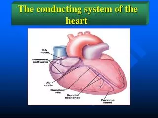
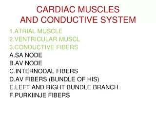
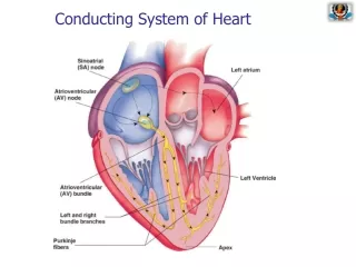
The heart beat is brought about by the activity of the pacemaker and the conducting system of the heart The pacemaker, known as the sino-atrial node (SAN) is located in the wall of the right atrium The region is composed of autorhythmic cells that exhibit spontaneous excitation
Impulses generate currents that can be detected by an ECG A wave of excitation originating in the SAN spreads through the muscle cells in the wall of the two atria This makes them contract simultaneously, atrial systole The impulse is then picked up by the atrio-ventricular node (AVN) located near the base of the atria The impulse passes from the AVN into a bundle of conducting fibres Stimulation of conducting fibres in the ventricular walls causes the simultaneous contraction of the two ventricles, ventricular systole
Heart Rate The medulla of the brain regulates heart rate The cardio-accelerator centre sends nerve impulses via the sympathetic nerve to the heart An increase in the number of nerve impulses arriving at the pacemaker via the sympathetic nerve results in an increase in heart rate The cardio-inhibitor centre in the medulla sends its information via a parasympathetic nerve to the heart An increase in the number of impulses arriving at the ASN from the parasympathetic nerve results in a decrease in heart rate
Hormonal Control Under certain circumstances, such as stress or exercise, the sympathetic nervous system acts on the adrenal glands making them release the hormone adrenaline (epinephrine) On reaching the SAN, the hormone makes the pacemaker generate cardiac impulses at a higher rate – bringing about an increase in heart rate
Electrocardiogram Electrical activity of the heart generates tiny currents that can be picked up on the skin surface The electrical signals produce a pattern called an electrocardiogram (ECG) The normal ECG consists of three distinct waves, normally referred to as P, QRS and T P corresponds to the wave of electrical excitation spreading over the atria from the pacemaker QRS represents the wave of excitation through the ventricles T corresponds to the electrical recovery of the ventricles at the end of ventricular systole
QRS, wave of excitation through ventricles P, electrical excitation over atria T, electrical recovery of ventricles
Blood Pressure Blood pressure is the force exerted by blood against the walls of blood vessels It is generated by the contraction of ventricles The highest value is in the Aorta and Pulmonary Artery During ventricular systole the blood pressure in the Aorta rises to 120mm Hg During ventricular diastole it drops to about 80mm Hg There is a progressive decrease in pressure as blood travels round the circulatory system – almost zero when it reaches the right atrium again Sphygmomanometer, below, measures systolic and diastolic pressure
Sphygmomanometer use Step 1 - the cuff is inflated until the pressure it exerts stops the blood flowing through the arm artery Step 2 - cuff allowed to deflate until pressure in artery exceeds pressure in cuff Blood can be heard spurting through artery and pulse can be felt The pressure at this stage is a measure of Systolic Pressure Step 3 - more air is released until spurting blood and pulse cannot be heard, this is a measure of the Diastolic Pressure
Hypertension Hypertension is prolonged elevation of the blood pressure when at rest High blood pressure usually involves values >140/>90 mm Hg Hypertension is a major risk for Coronary Heart Disease and Stokes It is commonly found in people with unhealthy lifestyles Overweight Not enough exercise Fatty diet Too much salt Excessive alcohol Continuous stress
Now try these questions . . . . 1. What brings about the heart beat? 2. What makes the atria contract? 3. What causes ventricular systole? 4. What part of the brain regulates heart rate? 5. What effect does adrenaline have on the heart rate? 6. What is blood pressure? 7. What is hypertension? 8. Name some common causes of hypertension
1. What brings about the heart beat? The heart beat is brought about by the activity of the pacemaker and the conducting system of the heart 2. What makes the atria contract? A wave of excitation originating in the SAN spreads through the muscle cells in the wall of the two atria This makes them contract simultaneously, atrial systole 3. What causes ventricular systole? Stimulation of conducting fibres in the ventricular walls causes the simultaneous contraction of the two ventricles, ventricular systole 4. What part of the brain regulates heart rate? The medulla of the brain regulates heart rate 5. What effect does adrenaline have on the heart rate? On reaching the SAN, the hormone makes the pacemaker generate cardiac impulses at a higher rate – bringing about an increase in heart rate
6. What is blood pressure? Blood pressure is the force exerted by blood against the walls of blood vessels, it is generated by the contraction of ventricles - the highest value is in the Aorta and Pulmonary Artery 7. What is hypertension? Hypertension is prolonged elevation of the blood pressure when at rest High blood pressure usually involves values >140/>90 mm Hg Hypertension is a major risk for Coronary Heart Disease and Stokes 8. Name some common causes of hypertension Being overweight Not enough exercise Fatty diet Too much salt Excessive alcohol Continuous stress