Download

1 / 94
950 likes | 1.42k Views
The Latest In Corneal Degenerations and Dystrophies. Blair B Lonsberry, MS, OD, MEd., FAAO Diplomate, American Board of Optometry Clinic Director and Professor Pacific University College of Optometry Portland, OR blonsberry@pacificu.edu. CORNEAL DYSTROPHIES. 2. 2. Corneal Dystrophies.

E N D
The Latest In Corneal Degenerations and Dystrophies Blair B Lonsberry, MS, OD, MEd., FAAO Diplomate, American Board of Optometry Clinic Director and Professor Pacific University College of Optometry Portland, OR blonsberry@pacificu.edu
Corneal Dystrophies • Group of corneal diseases that are: • genetically determined and • have been traditionally classified with respect to the corneal layer affected • Emerging molecular science: • is redefining traditional thought on the dystrophies and • offering potential avenues for therapeutic intervention.
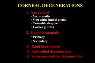
CORNEAL DEGENERATION • Non-familial, late onset • Asymmetric, unilateral, central or peripheral • Changes to the tissue caused by inflammation, age, or systemic disease. • Characterized by a deposition of material, a thinning of tissue, or vascularization
Epithelial (Anterior) Basement Membrane Dystrophy (EBMD or ABMD) • Primary features of this “dystrophy” are: • abnormal corneal epithelial regeneration and maturation, • abnormal basement membrane • Often considered the most common dystrophy, but may actually be an age-related degeneration. • large number of patients with this condition, • increasing prevalence with increasing age, and • its late onset support a degeneration vs. dystrophy.
Epithelial (Anterior) Basement Membrane Dystrophy (EBMD or ABMD) • Not all patients are symptomatic (range 10-69%) • Most common symptom is mild FB sensation which is worse in dry weather, wind and air conditioning • Blurred vision from irregular astigmatism or rapid TBUT • Pain is usually secondary to a RCE (recurrent corneal erosion) in apprx 10%
Epithelial (Anterior) Basement Membrane Dystrophy (EBMD or ABMD) • Easy to overlook: • typically bilateral though often asymmetric, • females>males, • often first diagnosed b/w ages of 40-70
Epithelial (Anterior) Basement Membrane Dystrophy (EBMD or ABMD) • Most common findings are: • chalky patches, • intraepithelial microcysts, and • fine lines (or any combination) in the central 2/3rd of cornea 8
Epithelial (Anterior) Basement Membrane Dystrophy (EBMD or ABMD) • Often referred to as: • maps, • dots or • fingerprints 9
Epithelial (Anterior) Basement Membrane Dystrophy (EBMD or ABMD): Treatment • Typically directed towards preventing RCE • If RCE’s develop: • awake with painful eye that improves as day wears on • chalky patches/dots in lower 2/3rd of cornea
RCE: Treatment • Initial treatment includes: • use of hyperosmotic ointment at bedtime, • bandage contact lens and • lubrication. 12
Recurrent Corneal Erosion: Treatment • If severe enough to cause vision loss or repeated episodes: • oral doxycycline with/without topical corticosteroid • Doxy 50 mg bid and FML tid for 4-8 weeks • both meds inhibit key metalloproteinases important in disease pathogenesis • Azasite (topical azithromycin) • debridement, • stromal puncture, or • PTK • Latest development: amniotic membrane transplant e.g. Prokera
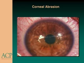
CORNEAL DEBRIDEMENT • Soften epithelium • 1-2 gtt topical anesthetic • q 15-30 seconds for 2-3 minutes • Use cotton swab, spatula, spud • or jewelers forceps • Remove flaps by pulling edges toward center • Don’t pull directly up or out • Remove flaps down to tight, • firm edges. • Tx abrasion (>50-100%) • Recurrence Rate 18%
Amniotic Membrane Transplant • Amniotic membrane is a biologic tissue with: • antiangiogenic, • antiscarring, • antimicrobial, and • anti-inflammatory properties that promotes healing of the ocular surface • Amniotic membrane grafts have been used for a variety of ocular conditions including: • Corneal burns • Neurotrophic ulcers • Stem cell damage • Persistent epithelial defects
Amniotic Membrane Transplant • Traditionally, amniotic membrane grafts had to be sutured • With the advent of tissue adhesives, amniotic transplants can now be sutureless • ProKera was approved by the FDA in 2003 as a Class II medical device which has a polycarbonate ring which holds a cryopreserved amniotic membrane • ProKera is indicated in the treatment of corneal erosions, neurotrophic corneas, recalcitrant corneal inflammation, acute ocular surface burns, acute Stevens Johnson syndrome, and descemetoceles.
RCE and LASIK • Patients who have a history of EBMD may not be ideal candidates for LASIK and should be carefully screened for prior to surgery. 18
Macular (Groenouw Type II) • Grayish opacities in the superficial stroma • With age: • extension into deeper stromal layers • intervening stroma becomes hazy • progressive loss of vision, • photophobia and ocular discomfort.
Macular Corneal Dystrophy • Surgical treatment usually required by 2nd or 3rd decade of life. • PK • DALK not indicated as may have damage to Descemets
Granular Dystrophy: (Groenouw Type I) • Discrete white granular opacities in central anterior corneal stroma. • With age: • increasing number, density, size and depth of opacities • intervening stroma and peripheral cornea remain clear
Granular Dystrophy: (Groenouw Type I) • RCE are common with associated pain. • Decreased vision results from subepithelial scarring or dense stromal deposits. • Surgical treatment includes penetrating keratoplasty or DALK (Deep Anterior Lamellar Keratoplasty). 22
PTK Treatment for GRANULAR SCLAFANI
Lattice (Type I) • Characteristic clinical appearance includes: • linear, • refractile, • branching deposits within the anterior stroma.
Lattice (Type I) • The central cornea is progressively opacified resulting is scarring and deterioration of vision while the periphery remains clear. • RCE’s often present. • May require surgical intervention with diminished vision. • PK • DALK 25
Central Crystalline Dystrophy of Schnyder • Opacities consist of: • small, needle-shaped refractile crystals that are either white or polychromatic • may extend into deeper stroma but epithelium remains normal. 26
Central Crystalline Dystrophy of Schnyder • Vision is typically mildly affected though there maybe associated systemic complications • systemic cholesterol should be evaluated
PK Surgery: Full Thickness Surgery Donor tissue sutured into recipient Recipient tissue removed Central trephine cut made Sutures create an irregular surface with astigmatism and blurring Full thickness block of tissue removed just to get to the endothelium Smooth Surface with only endothelial disease
Deep Anterior Lamellar Keratoplasty (DALK) • Removal of all tissue EXCEPT Descemet’s and endothelium • Most common rejection seen in PK is endothelial rejection observed in apprx 20% of low-risk cases • Repeated PK’s increase chance that the graft will be rejected • DALK can avoid risk of endothelial rejection with similar optical results as PK
Deep Anterior Lamellar Keratoplasty (DALK) • Indicated for patients with • Keratoconus • Corneal scars • Corneal stromal dystrophies • Basically any pathology that spares the endothelium • Contraindicated • Bullous keratopathy • Fuch’s
Deep Anterior Lamellar Keratoplasty (DALK) • Advantages over PK: • No “open sky” during surgery so lesser chance of expulsive hemorrhage • Much decreased rejection potential because patient keeps their own endothelium • Stromal rejection is rare and easily treated • Low to no rejection risk so steroids are tapered more quickly (usually twice as fast) • Heals faster as steroids tapered sooner allowing sutures to be removed earlier and more rapid visual stabilization (apprx 6 months) • More tectonic stability as patient keeps own endo
Normal Changes to the Endothelium • Descemet’s layer thickens from 3-17u • There is a decrease in the # of endothelial cells • from 3500 cells/mm2 to 1200 • this single layer spreads out: lacks mitosis • High density mitochondria : 90% pump • Lenses produce reversible polymegathism SCLAFANI
Abnormal Changes to the Endothelium • Endothelial cells become more irregular • Cells secrete collagen towards Descemet’s causing multilamination = guttata • This breaks down the barrier function and results in stromal and epithelial edema SCLAFANI
Fuch’s Dystrophy • Endothelium: • acts as both a barrier and pump function • responsible for maintaining corneal transparency by reducing corneal hydration • Fuch’s: • occurs bilaterally, • AD inheritance, • females 3x more likely to develop condition
Fuch’s Dystrophy: Guttata • Corneal guttata • excessive accumulation of abnormal endo secretions is associated with the disease process • usually first noticed in the central cornea in the patients 30’s and 40’s • corneal physiology is affected adversely by interference with pump action • guttata appear as small refractile “drops” on the corneal endo
Fuch’s Dystrophy: Guttata • closer inspection with specular reflection reveals an “orange peel-like” dimpling of the endo • with the decreased pump function, the overlying stroma becomes edematous • long standing corneal edema may result in corneal scarring and RCE
BS – Pre-Op Fuch’s Dystrophy Endothelial Cell Count: 545 cells/mm
Fuch’s Dystrophy • Patient symptoms vary with degree of guttata and compromised pump function • Moderate guttata • may affect visual function • may result in light scatter (haloes) • typically noticed upon waking • With increased disruption to the pump: • vision decreases • potential development of bullous keratopathy
Healthy endo: Cornea Thin and clear Endo dropout: Cornea swells, mild vision loss Severe swelling, blisters on surface, Va drops, pain Chronic swelling, surface scarring Stages of Fuch’s Dystrophy
Fuch’s Dystrophy: Treatment • Treatment in early stages: • usually palliative with the goal of improving comfort and function • hyperosmotics at bedtime (e.g. muro 128 ointment) may help reduce epithelial corneal edema in the morning • bandage CL can be used in the presence of bullous keratopathy
Fuch’s Dystrophy: Treatment • When visual function deteriorates to the point patient is unduly affected, surgical options are considered including: • penetrating keratoplasty (PK) • DLEK surgery (deep lamellar endothelial keratoplasty) or • newer DSAEK (Descemet Stripping Automated Endothelial Keratoplasty) • Latest DMEK (Descemet Membrane Endothelial Keratoplasty) • Fuch’s is leading reason for PK’s in developed countries 47 47
DLEK • Recipient cornea is stripped of its Descemet’s membrane, endothelium and posterior stroma • There is transplantation of the posterior stroma and endothelium of the donor cornea through a small incision • Results in improved: • endothelial function, • corneal clarity and • restoring useful vision.
DLEK • Procedure has: • minimal affect on refraction, • provides rapid visual recovery and • maintains structural integrity of the cornea. 49
DLEK Surgery: Split Thickness Surgery to replace only the diseased tissue Recipient tissue removed Scleral incision, deep corneal pocket, and endothelium trephined with Terry Trephine or cut with Cindy Scissors Just endothelium on posterior stromal disc removed from pocket Donor tissue placed into recipient Endothelium replaced with no sutures, supported by air bubble in anterior chamber. Surface remains smooth with no astigmatism