Download
1 / 53
1.11k likes | 2.44k Views
Thyroid Disorders. Khalid Al-Shali MBBS, MSc, FRCP(C), FACP Assistant Professor, Department of Medicine. Thyroid disorders: Hypothyroidism Hyperthyroidism and thyrotoxicosis Graves’ disease Thyroiditis Toxic adenoma Toxic multinodular goitre Thyrotoxicosis factitia Struma ovarii

E N D
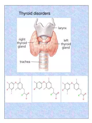
Thyroid Disorders Khalid Al-Shali MBBS, MSc, FRCP(C), FACP Assistant Professor, Department of Medicine
Thyroid disorders: Hypothyroidism Hyperthyroidism and thyrotoxicosis Graves’ disease Thyroiditis Toxic adenoma Toxic multinodular goitre Thyrotoxicosis factitia Struma ovarii Hydatidiform mole TSH-secreting pituitary adenoma Nontoxic goitre Thyroid nodules & thyroid cancer Benign thyroid nodules Thyroid cancer Papillary carcinoma Follicular carcinoma Medullary carcinoma Anaplastic carcinoma Lymphoma Cancer metastatic to the thyroid Introduction
Hypothyroidism • Etiology: • Primary: • Hashimoto’s thyroiditis with or without goitre • Radioactive iodine therapy for Graves’ disease • Subtotal thyroidectomy for Graves’ disease or nodular goitre • Excessive iodine intake • Subacute thyroiditis • Rare causes • Iodide deficiency • Goitrogens such as lithium; antithyroid drug therapy • Inborn errors of thyroid hormone synthesis • Secondary: Hypopituitarism • Tertiary: Hypothalamic dysfunction (rare) • Peripheral resistance to the action of thyroid hormone
Clinical features Cardiovascular signs: Bradycardia Low voltage ECG Pericardial effusion Cardiomegaly Hyperlipidemia Constipation, ascites Weight gain Cold intolerance Rough, dry skin Puffy face and hands Hoarse, husky voice Yellowish color of skin due to reduced conversion of carotene to vitamin A Respiratory failure Menorrhagia, infertility, hyper- prolactinemia Renal function: Impaired ability to excrete a water load Anemia: Impaired Hb synthesis Fe deficiency due to: Menorrhagia Reduced intestinal absorption Folate def. due to impaired intestinal absorption Pernicious anemia Neuromuscular system: Muscle cramps, myotonia Slow reflexes Carpal tunnel syndrome CNS symptoms: Fatigue, lethargy, depression Inability to concentrate Hypothyroidism
Hypothyroidism • Diagnosis: • A iFT4 and hTSH is diagnostic of primary hypothyroidism • Serum T3 levels are variable (maybe in normal range) • +ve test for thyroid autoantibodies (Tg Ab & TPO Ab) PLUS an enlarged thyroid gland suggest Hashimoto’s thyroiditis • With pituitary myxedema FT4 will be i but serum TSH will be inappropriately normal or low • TRH test may be done to differentiate pituitary from hypothalamic disease. Absence of TSH response to TRH indicates pituitary deficiency • MRI of brain is indicated if pituitary or hypothalamic disease is suspected. Need to look for other pituitary deficiencies. • If TSH is h & FT4 & FT3 are normal we call this condition subclinical hypothyroidism
Hashimoto’s Thyroiditis • Hashimoto’s thyroiditis is a commom cause of hypothyroidism and goitre especially in children and young adults. • It is an autoimmune disease that involves heavy infiltration of lymphocytes that totally destroys normal thyroidal architecture • Three different autoantibodies are present: Tg Ab, TPO Ab, and TSH-R Ab (block) • It is familial and may be associated with other autoimmune diseases such as pernicious anemia, adrenocortical insufficiency, idiopathic hypoparathyroidism, and vitiligo. • Shmidt’s syndrome consists of Hashimoto’s thyroiditis, adrenal insufficiency, hypoparathyroidism, DM, ovarian failure, and (rarely) candidal infections.
Hashimoto’s Thyroiditis • Symptoms & Signs: • Usually presents with goitre in a patient who is euthyroid or has mild hypothyroidism • Sex distribution: four females to one male • The process is painless • Older patients may present with severe hypothyroidism with only a small, firm atrophic thyroid gland • Transient symptoms of thyrotoxicosis can occur during periods of hashitoxicosis (spontaneously resolving hyperthyroidism) • Lab: • Normal or low thyroid hormone levels, and if low, TSH is elevated • High Tg Ab and/or TPO Ab titres • FNA bx reveals a large infiltration of lymphocytes PLUS Hurthle cells • Complications: • Permanent hypothyroidism (occurs in 10-15% of young pts) • Rarely, thyroid lymphoma
Management of Hypothyroidism • Start patient on L-thyroxine 0.05-0.1mg PO OD. L-thyroxine treats the hypothyroidism and leads to regression of goitre. • If patient is elderly or has IHD start 0.025mg PO OD. • Check TSH level after 4-6 weeks to adjust the dose of L-thyroxine. • In case of secondary hypothyroidism monitor FT4 instead of TSH. • Hypothyroidism during pregnancy: • Check TFT every month. L-thyroxine dose requirement tends to go up as the pregnancy progresses. • If patient has concommitant hyperprolactinemia and hypercholesterolemia, treat if not normalized after adequate thyroid replacement.
Myxedema Coma • Medical emergency, end stage of untreated hypothyroidism • Characterized by progressive weakness, stupor, hypothermia, hypoventilation, hypoglycemia, hyponatremia, shock, and death • The patient (or a family member) may recall previous thyroid disease, radioiodine therapy, or thyroidectomy • Hx is of gradual onset of lethargy progressing to stupor or coma. A hx of amenorrhea or impotence with pituitary myxedema • PE reveals iHR and marked hypothermia (as low as 24C) • The pt is usually an obese elderly woman with yellowish skin, a hoarse voice, a large tongue, thin hair, puffy eyes, ileus, and slow reflexes. An anterior neck scar may be present. Scanty pubic or axillary hair with pituitary myxedema • Lab: low FT4, TSH high, normal, or low, cholesterol high or N, serum Na low • ECG: bradycardia and low voltage • May be ppt by HF, pneumonia, excessive fluid administration, narcotics
Management of Myxedema Coma • Initiate therapy if presumptive clinical diagnosis after TSH, FT3 FT4 drawn. Also draw serum cortisol, ACTH, glucose. • General measures: • Patient should be in ICU setting • Support ventilation as respiratory failure is the major cause of death in myxedema coma • monitors ABG`s • support blood pressure; hypotension may respond poorly to pressor agents until thyroid hormone is replaced • hypothermia will respond to thyroxin therapy ; in interim use passivewarming only • hyponatremia will also be corrected by thyroxine therapy in majority of cases • hypoglycemia requires IV glucose • avoid fluid overload
Management of Myxedema Coma • Specific measure: • L-thyroxine 0.2-0.5 mg IV bolus, followed by 0.1 mg IV OD until oral therapy is tolerated • Results in clinical response in hours • Adrenal insufficiency may be precipitated by administration of thyroid hormone therefore hydrocortisone 100 mg IV q 8h is usually given until the results of the initial plasma cortisol is known. • Identify and treat the underlying precipitant cause
Graves’ Disease • Most common form of thyrotoxicosis • May occur at any age but mostly from 20-40 • 5 times more common in females than in males • Syndrome consists of one or more of the following: • Thyrotoxicosis • Goitre • Opthalmopathy (exopthalmos) and • Dermopathy (pretibial myxedema) • It is an autoimmune disease of unknown cause • 15% of pts with Graves’ have a close relative with the same disorder
Graves’ Disease • Pathogenesis: • T lymphocytes become sensitized to Ag within the thyroid gland and stimulate B lymphocytes to synthesize Ab to these Ag • One such Ab is the TSH-R Ab(stim), which stimulates thyroid cell growth and function • Graves’ may be ppt by pregnancy, iodide excess, viral or bacterial infections, lithium therapy, glucocorticoid withdrawal • The opthalmopathy and dermopathy associated with Graves’ may involve lymphocyte cytokine stimulation of fibroblasts in these locations causing an inflammatory response that leads to edema, lymphocytic infiltration, and glycosaminoglycans deposition • The tachycardia, tremor, sweating, lid lag, and stare in Graves’ is due to hyperreactivity to catecholamines and not due to increased levels of circulating catecholamines
Graves’ Disease • Clinical features: • I Eye features: Classes 0-6, mnemonic “NO SPECS” • Class 0: No signs or symptoms • Class 1: Only signs (lid retraction, stare, lid lag), no symptoms • Class 2: Soft tissue involvement (periorbital edema, congestion or redness of the conjunctiva, and chemosis) • Class 3: Proptosis (measured with Hertel exopthalmometer) • Class 4: Extraocular muscle involvement • Class 5: Corneal involvement • Class 6: Sight loss (optic nerve involvement)
Graves’ Disease • Clinical features: • II Goitre: • Diffuse enlargement of thyroid • Bruit may be present • III Thyroid dermopathy (pretibial myxedema): • Thickening of the skin especially over the lower tibia • The dermopathy may involve the entire leg and may extend onto the feet • Skin cannot be picked up between the fingers • Rare, occurs in 2-3% of patients • Usually associated with opthalmopathy and very hTSH-R Ab
Clinical features: IV Heat intolerance V Cardiovascular: Palpitation, Atrial fibrillation CHF, dyspnea, angina VI Gastrointestinal: Weight loss, happetite Diarrhea VII Reproductive: amenorrhea, oligo- menorrhea, infertility Gynecomastia VIII Bone: Osteoporosis Thyroid acropachy IX Neuromuscular: Nervousness, tremor Emotional lability Proximal myopathy Myasthenia gravis Hyper-reflexia, clonus Periodic hypokalemic paralysis X Skin: Pruritus Onycholysis Vitiligo, hair thinning Palmar erythema Spider nevi Graves’ Disease
Graves’ Disease • Diagnosis: • Low TSH, High FT4 and/or FT3 • If eye signs are present, the diagnosis of Graves’ disease can be made without further tests • If eye signs are absent and the patient is hyperthyroid with or without a goitre, a radioiodine uptake test should be done. • Radioiodine uptake and scan: • Scan shows diffuse uptake • Uptake is increased • TSH-R Ab (stim) is specific for Graves’ disease. May be a useful diagnostic test in the “apathetic” hyperthyroid patient or in the pt who presents with unilateral exopthalmos without obvious signs or laboratory manifestations of Graves’ disease
Treatment of Grave’s Disease • There are 3 treatment options: • Medical therapy • Surgical therapy • Radioactive iodine therapy
Treatment of Grave’s Disease • A. Medical therapy: • Antithyroid drug therapy: • Most useful in patients with small glands and mild disease • Treatment is usually continued for 12-18 months • Relapse occurs in 50% of cases • There are 2 drugs: • Neomercazole (methimazole or carbimazole): start 30-40mg/D for 1-2m then reduce to 5-20mg/D. • Propylthiouracil (PTU): start 100-150mg every 6hrs for 1-2m then reduce to 50-200 once or twice a day • Monitor therapy with fT4 and TSH • S.E.: 5%rash, 0.5%agranulocytosis (fever, sore throat), rare: cholestatic jaundice, hepatocellular toxicity, angioneurotic edema, acute arthralgia
Management of Grave’s disease • A. Medical therapy: • Propranolol 10-40mg q6hrs to control tachycardia, hypertension and atrial fibrillation during acute phase of thyrotoxicosis. It is withdrawn gradually as thyroxine levels return to normal • Other drugs: • Ipodate sodium (1g OD): inhibits thyroid hormone synthesis and release and prevents conversion of T4 to T3 • Cholestyramine 4g TID lowers serum T4 by binding it in the gut
Management of Grave’s disease • B. Surgical therapy: • Subtotal thyroidectomy is the treatment of choice for patients with very large glands • The patient is prepared with antithyroid drugs until euthyroid (about 6 weeks). In addition 2 weeks before the operation patient is given SSKI 5 drops BID to diminish vascularity of thyroid gland • Complications (1%): • Hypoparathyroidism • Recurrent laryngeal nerve injury
Management of Grave’s Disease • C. Radioactive iodine therapy: • Preferred treatment in most patients • Can be administered immediately except in: • Elderly patients • Patients with IHD or other medical problems • Severe thyrotoxicosis • Large glands >100g • In above cases it is desirable to achieve euthyroid state first • Hypothyroidism occurs in over 80% of cases. • Female should not get pregnant for 6-12m after RAI.
Management of Grave’s Disease • Management of opthalmopathy: • Management involves cooperation between the endocrinologist and the opthalmologist • A course of prednisone immediately after RAI therapy 100mg daily in divided doses for 7-14 days then on alternate days in gradually diminishing dosage for 6-12 weeks. • Keep head elevated at night to diminish periorbital edema • If steroid therapy is not effective external x-ray therapy to the retrobulbar area may be helpful • If vision is threatened orbital decompression can be used
Management of Grave’s Disease • Management during pregnancy: • RAI is contraindicated • PTU is preferred over neomercazole • FT4 is maintained in the upper limit of normal • PTU can be taken throughout pregnancy or if surgery is contemplated then subtotal thyroidectomy can be performed safely in second trimester • Breastfeeding is allowed with PTU as it is not concentrated in the milk
Toxic Adenoma (Plummer’s Disease) • This is a functioning thyroid adenoma • Typical pt is an older person (usually > 40) who has noted recent growth of a long-standing thyroid nodule • Thyrotoxic symptoms are present but no infiltrative opthalmopathy. PE reveals a nodule on one side • Lab: low TSH, high T3, slightly high T4 • Thyroid scan reveals “hot” nodule with suppressed uptake in contralateral lobe • Toxic adenomas are almost always follicular adenomas and almost never malignant • Treatment: same as for Grave’s disease
Toxic Multinodular Goitre • Usually occurs in older pts with long-standing MNG • PE reveals a MNG that may be small or quite large and may even extend substernally • RAI scan reveals multiple functioning nodules in the gland or patchy distribution of RAI • Hyperthyroidism in pts with MNG can often be ppt by iodide intake “jodbasedow phenomenon”. • Amiodarone can also ppt hyperthyroidism in pts with MNG • Treatment: Same as for Grave’s disease. Surgery is preferred.
Subacute Thyroiditis • Acute inflammatory disorder of the thyroid gland most likely due to viral infection. Usually resolves over weeks or months. • Symptoms & Signs: • Fever, malaise, and soreness in the neck • Initially, the patient may have symptoms of hyperthyroidism with palpitations, agitation, and sweat • PE: No opthalmopathy, Thyroid gland is exquisitely tender with no signs of local redness or heat suggestive of abscess formation • Signs of thyrotoxicosis like tachycardia and tremor may be present • Lab: • Initially, T4 & T3 are elevated and TSH is low, but as the disease progresses T4 & T3 will drop and TSH will rise • RAI uptake initially is low but as the pt recovers the uptake increases • ESR may be as high as 100. Thyroid Ab are usually not detectable in serum
Subacute Thyroiditis • Management: • In most cases only symptomatic Rx is necessary e.g. acetaminophen 0.5g four times daily • If pain, fever, and malaise are disabling a short course of NSAID or a glucocorticoid such as prednisone 20mg three times daily for 7-10 days may be necessary to reduce the inflammation • L-thyroxine is indicated during the hypothyroid phase of the illness. 10% of the patients will require L-thyroxine long term
Other Forms of Thyrotoxicosis • Thyrotoxicosis Factitia: • Due to ingestion of excessive amounts of thyroxine • RAI uptake is nil and serum thyroglobulin is low • Struma Ovarii: • Teratoma of the ovary with thyroid tissue that becomes hyperactive • No goitre or eye signs. RAI uptake in neck is nil but body scan reveals uptake of RAI in the pelvis. • Hydatidiform mole: • Chorionic gonadotropin is produced which has intrinsic TSH-like activity. • TSH-secreting pituitary adenoma: • FT4 & FT3 is elevated but TSH is normal or elevated • Visual field examination may reveal temporal defects, and CT or MRI of the sella usually reveals a pituitary tumour.
Thyroid storm (Thyrotoxic crisis) • Usually occurs in a severely hyperthyroid patient caused by a precipitating event such as: • Infection • Surgical stress • Stopping antithyroid medication in Graves’ disease • Clinical clues • fever hyperthermia • marked anxiety or agitation coma • Anorexia • tachycardia tachyarrhythmias • pulmonary edema/cardiac failure • hypotension shock • confusion
Thyroid storm (Thyrotoxic crisis) • Initiate prompt therapy after free T4, free T3, and TSH drawn without waiting for laboratory confirmation. • Therapy • 1. General measures: • Fluids, electrolytes and vasopressor agents should be used as indicated • A cooling blanket and acetaminophen can be used to treat the pyrexia • Propranolol for beta–adrenergic blockade and in addition causesdecreased peripheral conversion of T4T3 but watch for CHF. • The IV dose is 1 mg/min until adequate beta-blockade has been achieved. Concurrently, propranolol is given orally or via NG tube at a dose of 60 to 80 mg q4h
Thyroid storm (Thyrotoxic crisis) • Therapy • 2. Specific Measures: • PTU is the anti-thyroid drug of choice and is used in high doses: 1000 mg of PTU should be given p.o. or be crushed and given via nasogastric tube, followed by PTU 250mg p.o. q 6h. If PTU unavailable can give methimazole 30mg p.o. every 6 hours. • One hour after the loading dose of PTU is given –give iodide which acutely inhibits release of thyroid hormone, i.e. Lugol’s solution 2-3 drops q 8h OR potassium iodide (SSKI) 5 drops q 8h. • Dexamethasone 2 mg IV q 6h for the first 24-48 hours lowers body temperature and inhibits peripheral conversion of T4-T3 • With these measures the patient should improve dramatically in the first 24 hours. • 3. Identify and treat precipitating factor.
Nontoxic Goitre • Enlargement of the thyroid gland from TSH stimulation which in turn results from inadequate thyroid hormone synthesis • Etiology: • Iodine deficiency • Goitrogen in the diet • Hashimoto’s thyroiditis • Subacute thyroiditis • Inadequate hormone synthesis due to inherited defect in thyroidal enzymes necessary for T4 and T3 biosynthesis • Generalized resistance to thyroid hormone (rare) • Neoplasm, benign or malignant
Nontoxic Goitre • Symptoms and Signs: • Thyroid enlargement, diffuse or multinodular • Huge goitres may produce a positive Pemberton sign (facial flushing and dilation of cervical veins on lifting the arms over the head) especially when they extend inferiorly retrosternally • Pressure symptoms in the neck with upward or downward movement of the head • Difficulty swallowing, rarely vocal cord paralysis • Most pts are euthyroid but some are mildly hypothyroid • RAI uptake and scan: • Uptake may be normal, low, or high depending on the iodide pool • Scan reveals patchy uptake with focal areas of increased and decreased uptake corresponding to “hot” and “cold” nodules respectively
Management of Nontoxic Goitre • L-thyroxine suppressive therapy: • Doses of 0.1 to 0.2mg daily is required • Aim is to suppress TSH to 0.1-0.4 microU/L (N 0.5-5) • Suppression therapy works in 50% of cases if continued for 1 year • If suppression does not work or if there are obstructive symptoms from the start then surgery is necessary
Benign Thyroid Nodules • Thyroid nodules are common especially among older women • Etiology: • Focal thyroiditis • Dominant portion of multinodular goitre • Thyroid, parathyroid, or thyroglossal cysts • Agenesis of a thyroid lobe • Postsurgical remnant hyperplasia or scarring • Postradioiodine remnant hyperplasia • Benign adenomas: • Follicular • Colloid or macrofollicular • Hurthle cell • Embryonal • Rare: Teratoma, lipoma, hemangioma
Thyroid Cancer Approximate frequency of malignant thyroid tumours
Papillary Carcinoma • Usually presents as a nodule that is firm, solitary, “cold” on isotope scan, and usually solid on thyroid US • In MNG, the cancer is usually a “dominant nodule” that is larger, firmer and different from the rest of the gland • 10% of papillary ca present with enlarged cervical nodes • Grows very slowly and remains confined to the thyroid gland and local lymph nodes for many years. • In later stages they can spread to the lung • Death usually from local disease or lung metastases • May convert to undifferentiated carcinoma • Many of these tumours secrete thyroglobulin which can be used as a marker for recurrence or metastasis of the cancer
Follicular Carcinoma • Differs from follicular adenoma by the presence of capsular or vascular invasion • More aggressive than papillary ca and can spread either by local invasion of lymph nodes or by blood vessel invasion with distant metastases to bone or lung • Death is due to local extension or to distant bloodstream metastasis with extensive involvement of bone, lungs & viscera • These tumours often retain the ability to concentrate RAI • A variant of follicular carcinoma is the “Hurthle cell” carcinoma. These tumours behave like follicular cancer except that they rarely take up RAI • Thyroglobulin secretion by follicular carcinoma can be used to follow the course of the disease