Download
1 / 34
360 likes | 752 Views
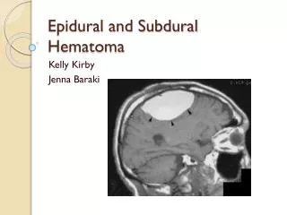
Once a vessel is torn, blood accumulating under arterial pressure can dissect the tightly applied dura away from the inner skull surface producing a hematoma that compresses the brain surface.

E N D
Once a vessel is torn, blood accumulating under arterial pressure can dissect the tightly applied dura away from the inner skull surface producing a hematoma that compresses the brain surface. - Clinically, patients can be lucid for several hours between the moment of trauma and the development of neurologic signs.. - An epidural hematoma may expand rapidly and constitutes a neurosurgical emergency necessitating prompt drainage and repair to prevent death
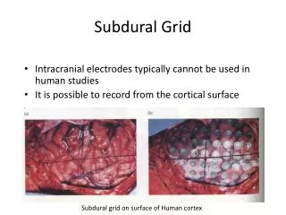
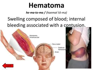
B. Subdural Hematoma :Mechanism - Rapid movement of the brain during trauma can tear the bridging veins that extend from the cerebral hemispheres through the subarachnoid and subdural space to the dural sinuses. • Their disruption produces bleeding into the subdural space • and the Susceptible persons are:
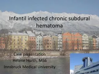
1. Patients with brain atrophy, the bridging veins are stretched out, and the brain has additional space within which to move, accounting for the higher rate of subdural hematomas in elderly persons. 2. Infants also are susceptible to subdural hematomas because their bridging veins are thin-walled.
- Subdural hematomas typically become manifest within the first 48 hours after injury. - They are most common over the lateral aspects of the cerebral hemispheres and may be bilateral. • Neurologic signs are attributable to the pressure exerted on the adjacent brain. - Symptoms may be localizing but more often are nonlocalizing, taking the form of headache
, confusion, and slowly progressive neurologic deterioration. MORPHOLOGY - An acute subdural hematoma appears as a collection of freshly clotted blood apposed to the contour of the brain surface, without extension into the depths of sulci. • The underlying brain is flattened, and the subarachnoid space is often clear - Typically, venous bleeding is self-limited; breakdown and organization of the hematoma take place over time
. Subdural hematomas organize by: 1. Lysis of the clot (about 1 week), 2. Growth of granulation tissue from the dural surface into the hematoma (2 weeks) 3. Fibrosis (1 to 3 months) - Organized hematomas are attached to the dura , but not to the underlying arachnoid - Fibrosing lesions may eventually retract, leaving only a thin layer of connective tissue ("subdural membranes").
- Subdural hematomas commonly rebleed (resulting in chronic subdural hematomas), presumably from the thin-walled vessels of the granulation tissue, leading to microscopic findings consistent with hemorrhages of varying age. - Symptomatic subdural hematomas are treated by surgical removal of the blood and associated reactive tissue
Dysmyelinating diseases,:Leukodystrophies - Are Inherited diseases in which the clinical symptoms derive from the abnormal myelin synthesis or turnover and some of these disorders. 1. Involve lysosomalenzymes or peroxisomal enzymes; • A few are associated with mutations in myelin proteins. - Most are of autosomal recessive inheritance, although X-linked diseases also occur
MORPHOLOGY 1. The white matter is diffusely gray and translucent with decreased volume. 2. Early in their course, some diseases may show patchy involvement, while others have a predilection for occipital lobe involvement. 3. In the end,nearly all of the white matter usually is affected.andthe brain becomes atrophic with enlarged ventricles 5. Myelin loss leads to infiltration of macrophages, which often become stuffed with lipid.
Clinical Features • Each of the various leukodystrophies has a characteristic clinical presentation, and most can be diagnosed by genetic or biochemical methods • Affected children are normal at birth but begin to miss developmental milestones during infancy and childhood • Diffuse involvement of white matter leads to deterioration in motor skills, spasticity, or ataxia.
II. Demyelinating diseases of the CNS - Are acquired conditions characterized by preferential damage to previously normal myelin and include: 1. Immune-mediated injury, such as multiple sclerosis (MS) 2. Progressive multifocal leukoencephalopathy 3. And injury caused by drugs and other toxic agents
1. Multliple sclerosis: - Is an autoimmune demyelinating disorder - It is the most common of the demyelinating disorders,. - The disease may become apparent at any age, although onset in childhood or after age 50 is relatively rare. - Women are affected twice as often as men
PATHOGENESIS : - A combination of environmental and genetic factors result in a loss of tolerance to self proteins (myelin antigens). - The nature of the initiating agent, often suggested to be an- infectious agent, remains uncertain. A- There is a significant contribution of genetic factors to the risk of developing MS 1.The disease risk is 15-fold higher when the disease is present in a first-degree relative
2. The concordance rate for monozygotic twins is about 25%, with a much lower rate for dizygotic twins 3. A significant fraction of the genetic risk for MS is attributable to HLA-DR variants and DR2 allele is the one that most significantly increases the risk for developing MS. 4. Polymorphisms in the genes encoding receptors for the cytokines IL-2 and IL-7 , which control the activation and regulation of T cell-mediated immune responses.
A. Pathophysilogy of loss of tolerance - Although myelin proteins are considered TSAs whose expression is largely restricted to the central nervous system, they are expressed within the thymus. • Among the myelin-specific T cells shown to escape central tolerance, most have been characterized as low avidity T cells that do not engage their antigen during maturation with sufficient strength to induce deletion. • These autoreactive T-cells against myelin antigen
will release cytokines the damage the blood brain barrier and causes the autommune reaction against myelin B. The mechanism of demyelination - Immune-mediated myelin destruction is thought to have a central role in MS exemplified by the central role for CD4+ T cells with an increase in TH17 and TH1 CD4+ cells thought to be a critical component of the injury to myelin. . - There is also contributions from CD8+ T cells and B cells
C. Mechanism of damage to axons - While MS is characterized by the presence of de-myelination out of proportion to axonal loss, some injury to axons does occur because chronic and recurrent inflammation release cytotoxic enzymes , cytokines ,matrix metalloproteases and NO from infiltrating lymphocytes cause damage to axons
Chronic multiple sclerosis patterns 1.Relapsing remitting pattern • Most common pattern • Usually symptoms have acute or subacute onset, with symptoms increasing over days to weeks followed by a period of a period of recovery over 1-2 months
- Remission of symptoms occurs due to redistribution of sodium channels throughout the axolemma of demylinated segment. • But overtime, over time there is usually a gradual, often stepwise, accumulation of neurologic deficits - Changes in cognitive function can be present, but are often much milder than the other deficits - In any individual patient, it is hard to predict when the next relapse will occur;
2. Secondary progressive MS • Second most common • Begins similar to the first pattern but evolves with an unremitting progressive course 3. Primary progressive MS - Have steadily progressive course from the onset
. MORPHOLOGY : - The affected areas show multiple, well-circumscribed, slightly depressed, gray irregularly shaped lesions termed plaques commonly arise near the ventricles - They are also frequent in • Optic nerves and chiasm, • Brain stem ascending and descending fiber tracts, c. Cerebellum, and spinal cord
In an active plaque : - There is evidence of ongoing myelin breakdown with macrophages containing myelin debris. - Lymphocytes and macrophages are present, mostly as perivascular cuff • Small active lesions often are centered on small veins • Axons are relatively preserved but reduced in number.
.B.Quiescent Inactive plaques: - Disappearance of inflammation leaving behind little to no myelin • Gliosis . C. Shadow plaques • Partial reduction in myelin density • Represent remyelinated areas
Mechanism of Remyelination • Remyelination Of demyelinated axons is necessary for restoring their function • But It is incomplete • It occurs due to migration of oligodendrocyte precursor cells to the lesions; therefore can proliferate and differentiate but are usually unable to completely remyelinate remaining axons
Failure of remyelination • Possible mechanisms • Reexpression on demyelinated axon surface in MS plaques of neural cell adhesion molecule (NCAM), a negative signal for myelination • Astrocytes may form dense glial scar both within the plaque and at its edge, so may prevent migration of oligodendocyte precursor cells to the lesions
Clinically isolated patterns • optic neuritis • Is the initial clinical event in20% of cases of MS • But may be clinically isolated • Symptoms include loss of visual activity, loss of peripheral vision or loss of colour vision • May be followed by optic atrophy
2. Neuromyelitisoptica (NMO)(Devic disease) : - Is an inflammatory demyelinating disease of the optic nerves and spinal cord and previously thought to be a form of MS with stereotypic anatomic regions of susceptibility - This is now recognized to be an antibody-mediated autoimmune disorder with antibodies to the water channel aquaporin-4 are both diagnostic and pathogenic.
- Imaging studies have demonstrated that there are often more lesions in the brains of patients with MS than might be expected from the clinical examination, • Lesions can come and go much more often than was previously suspected. - Most current treatments, which are intended to control the immune response, aim at decreasing the rate and severity of relapses rather than recovering lost function.
- The CSF shows: 1. A mildly elevated protein level with an increased proportion of immunoglobulin 2. In one third of cases, there is moderate pleocytosis. 3. When the immunoglobulin is examined further, oligoclonal bands usually are identified. • These antibodies are directed against a variety of antigens and can be used as markers of disease activity.
Other Acquired Demyelinating Diseases . 1. Central pontinemyelinolysis : - Is a nonimmune process - Characterized by loss of myelin involving the center of the pons but may affect other regions of the brain, most often after rapid correction of hyponatremia - The mechanism of oligodendroglial cell injury is uncertain, but it may be related to edema induced
by sudden changes in osmotic pressureIt occurs in a variety of clinical settings including alcoholism and severe electrolyte or osmolar imbalance. • Because of the involvement of fibers in the pons carrying signals to motor neurons in the spinal cord, patients often present with rapidly evolving quadriplegia. 2. Progressive multifocal leukoencephalopathy (PML) :Is a demyelinating disease that occurs after reactivation of JC virus in immunosuppressed patients.